. 2016 Feb;36(2):328-38.
doi: 10.1161/ATVBAHA.115.306091. Epub 2015 Dec 3.

Valve Endothelial Cell-Derived Tgfβ1 Signaling Promotes Nuclear Localization of Sox9 in Interstitial Cells Associated With Attenuated Calcification

Affiliations

Valve Endothelial Cell-Derived Tgfβ1 Signaling Promotes Nuclear Localization of Sox9 in Interstitial Cells Associated With Attenuated Calcification

Danielle J Huk et al. Arterioscler Thromb Vasc Biol. 2016 Feb.

Abstract

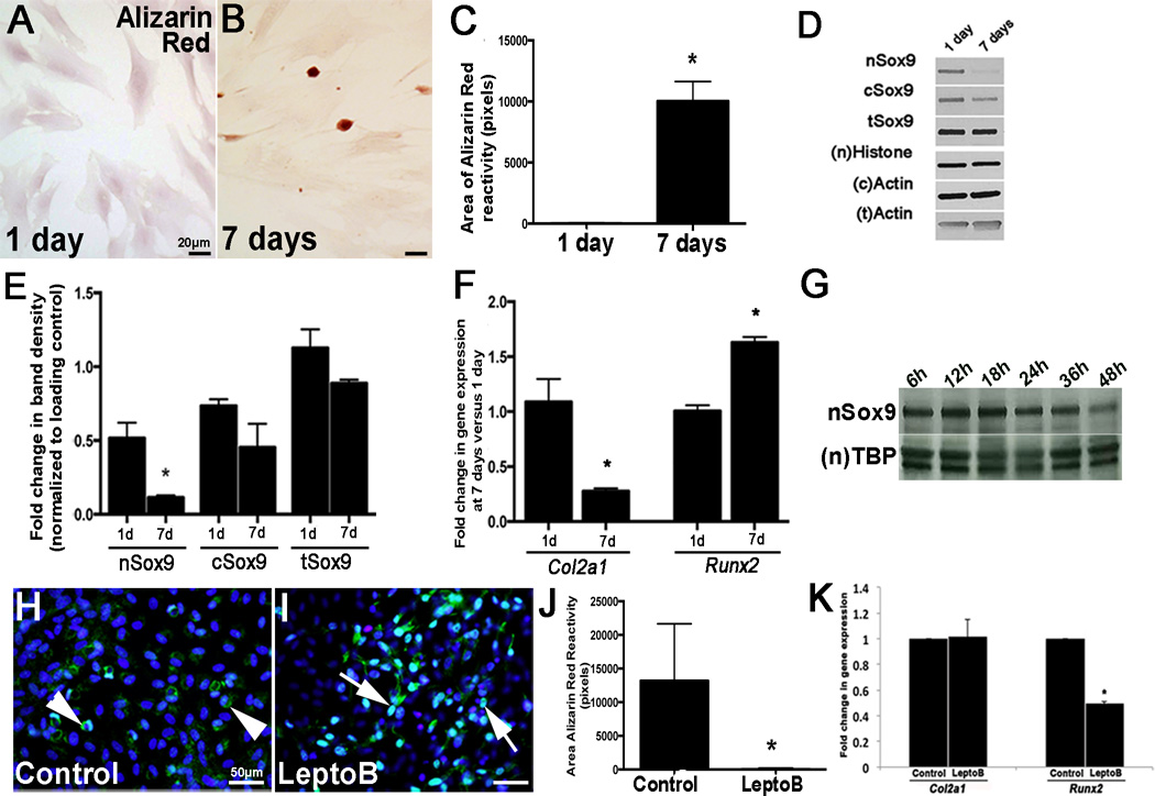
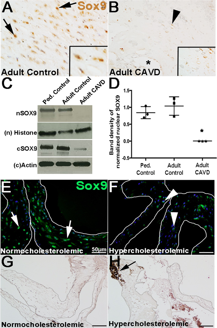
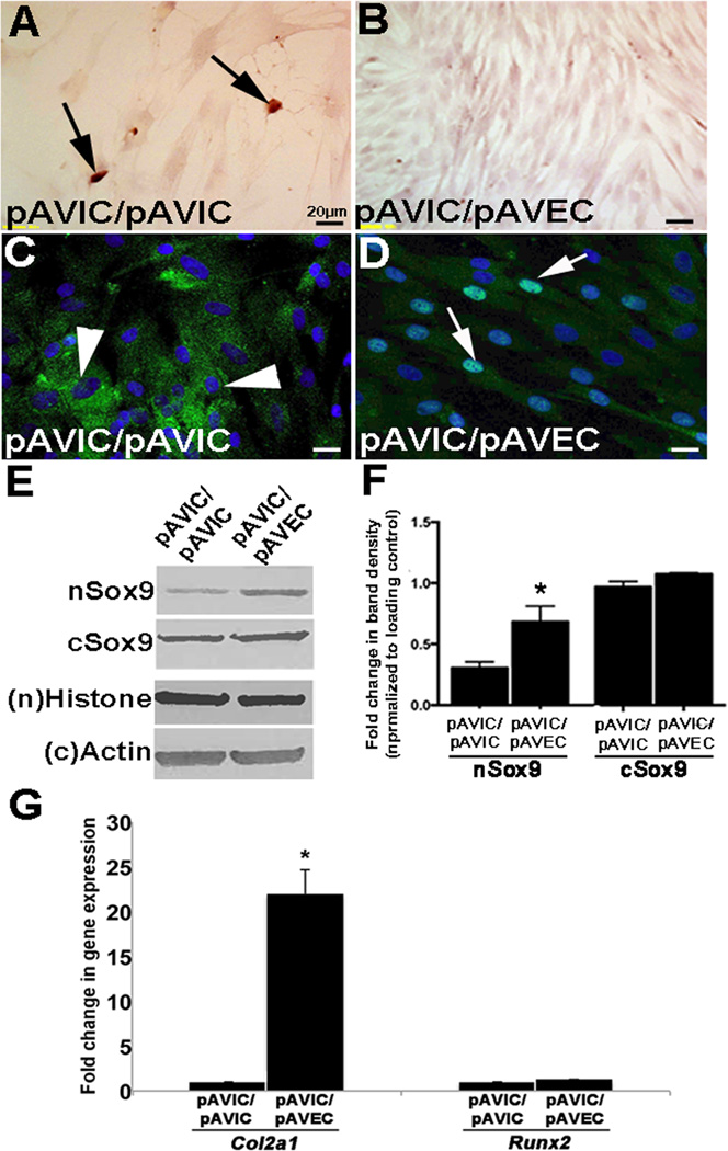
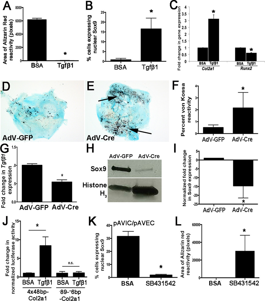
Objective: Aortic valve disease, including calcification, affects >2% of the human population and is caused by complex interactions between multiple risk factors, including genetic mutations, the environment, and biomechanics. At present, there are no effective treatments other than surgery, and this is because of the limited understanding of the mechanisms that underlie the condition. Previous work has shown that valve interstitial cells within the aortic valve cusps differentiate toward an osteoblast-like cell and deposit bone-like matrix that leads to leaflet stiffening and calcific aortic valve stenosis. However, the mechanisms that promote pathological phenotypes in valve interstitial cells are unknown.

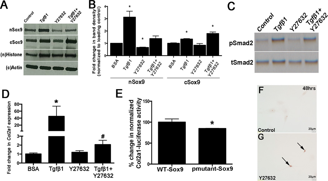
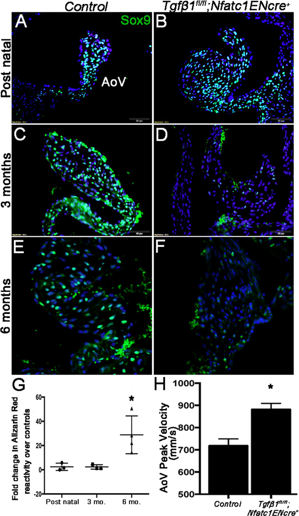
Approach and results: Using a combination of in vitro and in vivo tools with mouse, porcine, and human tissue, we show that in valve interstitial cells, reduced Sox9 expression and nuclear localization precedes the onset of calcification. In vitro, Sox9 nuclear export and calcific nodule formation is prevented by valve endothelial cells. However, in vivo, loss of Tgfβ1 in the endothelium leads to reduced Sox9 expression and calcific aortic valve disease.

Conclusions: Together, these findings suggest that reduced nuclear localization of Sox9 in valve interstitial cells is an early indicator of calcification, and therefore, pharmacological targeting to prevent nuclear export could serve as a novel therapeutic tool in the prevention of calcification and stenosis.

Keywords: animal model cardiovascular disease; aortic valve; endothelial cell; heart valve; signaling pathways.


Figures

Figure 1
Figure 1. Nuclear Sox9 localization is reduced in pAVIC calcification assays
(A, B) Alizarin Red staining to detect calcific nodule formation in pAVICs cultured for 1 (A) and 7 (B) days on glass. (C) Quantitation of Alizarin Red reactivity (in pixels, n=4). (D) Western blot analysis to show nuclear (n) and cytoplasmic (c) Sox9 in pAVICs cultured for 1, or 7 days. (E) Quantitation of Western blot shown in (D), normalized to respective loading controls. (F) qPCR to show changes in expression of Col2a1 and Runx2 in pAVICs cultured for 1, or 7 days. (G) Western blot analysis of nSox9 expression in pAVICs at the indicated time points. *, p<0.05 compared to 1 day cultures, n=3. (H, I) Immunohistochemistry to show Sox9 localization in control (H) or Leptomycin B treated pAVICs (I). (J) Quantitation of Alizarin Red positive nodule area. (K) qPCR to show Col2a1 and Runx2 expression in control and LeptomycinB-treated pAVICs. * p ≤ 0.05 compared to vehicle control.
Figure 2
Figure 2. Sox9 expression is reduced in calcified valves from human patients and mouse models
(A–B) Colormetric immunohistochemistry to detect Sox9 expression in VICs in AoV tissue sections isolated from non-diseased adult (~70 years of age) subjects (A) and age-matched control CAVD patients (B). Arrows indicate nuclear localization, arrowheads denote cytoplasmic expression, * shows calcific lesion. (C, D) Western blot (C) and quantitation (D, n=3) to show nuclear (n) and cytoplasmic (c) SOX9 expression in independent human samples collected from diseased, non-calcified pediatric controls (lane 1), non-diseased adult controls (lane 2) and age-matched control CAVD patients (lane 3). Immunofluorescence to detect Sox9 (green) expression (E, F) and Alizarin Red to stain calcific nodules (G, H) in AoVs isolated from Reversa hypercholesterolemic and normocholesterolemic control mice at 18 months of age. Arrow in E, F indicate nuclear localization, arrowheads denote cytoplasmic expression. Arrow in H shows calcific lesion. * p ≤ 0.05 compared to pediatric non-diseased controls.
Figure 3
Figure 3. Endothelial cells prevent VIC-mediated calcification and promote Sox9 nuclear localization
Alizarin Red staining to detect calcific nodules (arrows) (A, B) and immunohistochemistry to detect Sox9 expression and localization (C, D) in pAVICs cultured in the absence (A, C) or presence (B, D) of pAVECs, n=4. (E) Western blot analysis to show nuclear (n) and cytoplasmic (C) Sox9 in pAVICs co-cultured with pAVICs or pAVECs. (F) Quantitation of Western blot shown in (E), normalized to respective loading controls, based on n=3. (G) qPCR analysis of Col2a1 and Runx2 in pAVICs cultured in the absence and presence of pAVECs. *, p<0.05 compared to pAVIC/pAVIC experiments.
Figure 4
Figure 4. Tgfβ1 treatment prevents formation of calcific nodules by VICs and promotes Sox9 nuclear localization in VICs
(A) Quantitation of Alizarin Red staining to detect calcific nodule formation in pAVICs treated with BSA or 10ng/mL TGFβ1 for 7 days. (B) Quantitation of the number of pAVICs expressing nuclear Sox9 over the total number of cells for each treatment. (C) qPCR of Col2a1 and Runx2 expression in BSA and TGFβ1 treated cells.*, p<0.05 compared to BSA controls, based on n=4. (D, E) von Kossa staining of AoV explants isolated from Tgfβ1fl/fl neonate pups and treated with AdV-Cre (E) or AdV-GFP (D). von Kossa reactivity is quantified in (F). (G) qPCR to show decreased Tgfβ1fl expression in AoV explant assays and Western blot to show reduced Sox9 expression relative to AdV-GFP treated controls (H, I). (J) Luciferase assay to show transcriptional activity of Col2a1 in response to BSA and TGFβ1 treatment. 4×48bp-Col2a1 contains the Col2a1 minimal mouse promoter and Sox9-responsive enhancer region of intron 1. −89-+6bp-Col2a1 contains the minimal promoter only, n=3. (K, L) Quantitation of nuclear Sox9 localization and Alizarin Red reactivity in pAVICs following co-culture with pAVECs and treated with BSA, or the TGFβ inhibitor, SB43152. *, p<0.05 compared to BSA controls, based on n=3.
Figure 5
Figure 5. Tgfβ1-mediated regulation of Sox9 expression requires Rho kinase
Western blot analysis of nuclear (n) and cytoplasmic (c) Sox9 (A) and pSmad2 (C) expression in protein lysates collected from pAVICs treated with BSA (control), Tgfβ1, the ROCK inhibitor Y27632, or Tgfβ1 and Y27632. (B) Quantitation of A. (D) qPCR to detect changes in Col2a1 and Runx2 expression in treated cells. * p ≤ 0.05 compared to BSA control. (E) Luciferase assay to show Col2a1 (4×48bp-Col2a1) transcriptional activity in response to wild-type Sox9 (WT-Sox9) and a mutant Sox9 construct in which S64 and S181 phosphorylation sites have been abrogated (pmutant-Sox9). *, p<0.05 compared to WT-Sox9, based on n=3. (F, G) Alizarin Red staining to detect calcific nodule formation in pAVICs treated with BSA control or Y27632.
Figure 6
Figure 6. Targeted deletion of Tgfβ1 in murine VECs leads to decreased Sox9 expression, calcific nodule formation and AoV dysfunction in vivo
(A–F) Immunohistochemistry to show Sox9 expression (green, arrows) in AoV from control (Tgfβ1fl/fl;Nfatc1ENCre) (A, C, E) and Tgfβ1fl/fl;Nfatc1ENCre+ (B, D, F) mice at post natal (A, B), 3 (C, D) and 6 months (E, F) of age. Representation images shown based on n=3. (G) Alizarin Red reactivity in Tgfβ1fl/fl;Nfatc1ENCre+ mice at each time point relative to age-matched controls, n=3. (H) Echocardiography to determine AoV peak velocity in Tgfβ1fl/fl;Nfatc1ENCre+ mice at 6 months of age compared to controls, n=6. AoV, Aortic valve. *p<0.05 compared to controls.

References

    1. Go AS, Mozaffarian D, Roger VL, et al. American Heart Association Statistics C and Stroke Statistics S. Heart disease and stroke statistics--2014 update: a report from the American Heart Association. Circulation. 2014;129:e28–e292. - PMC - PubMed
    1. Beckmann E, Grau JB, Sainger R, Poggio P, Ferrari G. Insights into the use of biomarkers in calcific aortic valve disease. The Journal of heart valve disease. 2010;19:441–452. - PMC - PubMed
    1. Hinton RB, Jr, Lincoln J, Deutsch GH, Osinska H, Manning PB, Benson DW, Yutzey KE. Extracellular matrix remodeling and organization in developing and diseased aortic valves. Circulation research. 2006;98:1431–1438. - PubMed
    1. Freeman RV, Otto CM. Spectrum of calcific aortic valve disease: pathogenesis, disease progression, and treatment strategies. Circulation. 2005;111:3316–3326. - PubMed
    1. Gould ST, Matherly EE, Smith JN, Heistad DD, Anseth KS. The role of valvular endothelial cell paracrine signaling and matrix elasticity on valvular interstitial cell activation. Biomaterials. 2014;35:3596–3606. - PMC - PubMed

Publication types

MeSH terms

Supplementary concepts