. 2015 Dec 3:16:374.
doi: 10.1186/s12891-015-0827-4.

Back pain was less explained than leg pain: a cross-sectional study using magnetic resonance imaging in low back pain patients with and without radiculopathy

Affiliations

Back pain was less explained than leg pain: a cross-sectional study using magnetic resonance imaging in low back pain patients with and without radiculopathy

Ole Kudsk Jensen et al. BMC Musculoskelet Disord. .

Abstract

Background: Cross-sectional studies have shown associations between lumbar degenerative manifestations on magnetic resonance imaging (MRI) and low back pain (LBP). Disc herniations and other degenerative manifestations, however, frequently occur in asymptomatic individuals. The purpose of this cross-sectional study was to analyze for associations between pain intensity and degenerative manifestations and other pain variables in patients for whom prognostic factors have been published previously.

Methods: Included were 141 consecutive patients with and without radiculopathy, all sick-listed 1-4 months due to low back pain and subsequently examined by MRI of the lumbar spine. Using different methods of grouping the degenerative manifestations, linear regression analyses were performed with the intensity of back + leg pain, back pain and leg pain as dependent variables covering actual pain and pain the preceding 2 weeks. The clinical classification into +/- radiculopathy was established before and independently of the standardised description of MRI findings.

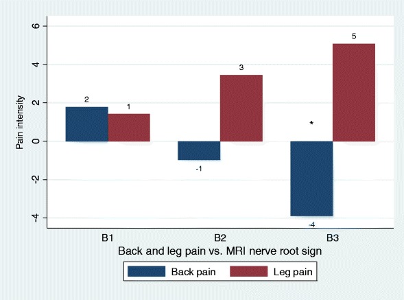
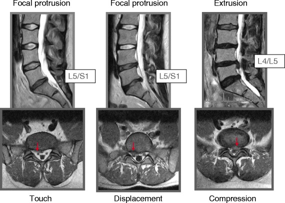
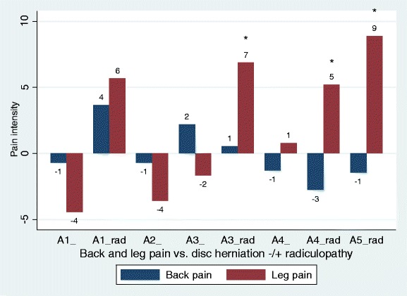
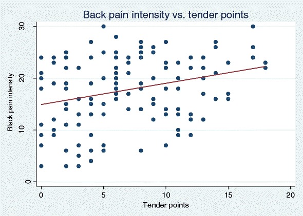
Results: Radiculopathy was present in 43 % of the patients. Pain was best explained using rank-ordered degenerative manifestations on MRI. Back pain and leg pain were differently associated, and back pain was less explained than leg pain in the multivariate analyses (15 % vs. 31 % of the variation). Back pain intensity was higher in patients with type 1 Modic changes and in some patients with nerve root touch, but was not associated with disc herniations. Leg pain intensity was well explained by disc herniations causing MRI nerve root compromise and radiculopathy. In patients with radiculopathy, nerve root touch caused as much leg pain as nerve root displacement or compression. High intensity zones and osteophytes were not associated with back pain, but only associated with leg pain in patients with radiculopathy. Tender points explained some of the back pain, and widespread pain explained leg pain in some of the patients without radiculopathy.

Conclusions: Back pain was associated with type 1 Modic changes, nerve root touch and tender points, whereas leg pain was associated with osteophytes, HIZ, disc herniation, all sorts of MRI nerve root compromise, radiculopathy and widespread pain.


Figures

Fig. 1
Fig. 1
Back pain and leg pain in relation to different types of disc herniations. Overall, only associations with leg pain were statistically significant (Table 2), statistical significance by type indicated by ‘*’. Negative values should be interpreted as less pain as compared to the reference group (no disc herniation). A1, bulging; A2, broad protrusion; A3, focal protrusion; A4, extrusion; A5, sequester
Fig. 2
Fig. 2
Back pain and leg pain in relation to different types of MRI nerve root sign. Overall, only associations with back pain were statistically significant (Table 2), statistical significance by type indicated by ‘*’. Negative values should be interpreted as less pain as compared to the reference group (no MRI nerve root sign). B1, nerve root touch; B2, nerve root displacement; B3, nerve root compression
Fig. 3
Fig. 3
Focal protrusion with nerve root touch (left) or displacement (middle) and extrusion with nerve root compression (right)
Fig. 4
Fig. 4
Back pain and leg pain in relation to different types of disc herniations with and without radiculopathy. Overall, only associations with leg pain were statistically significant (Table 3), statistical significance by type indicated by ‘*’. Negative values should be interpreted as less pain as compared to the reference group (no disc herniation). A1, bulging; A2, broad protrusion; A3, focal protrusion; A4, extrusion; A5, sequester, ‘rad’ indicating + radiculopathy. A2_rad and A5_ omitted as these subgroups included 1 and 0 patients, respectively
Fig. 5
Fig. 5
Back pain and leg pain in relation to different types of MRI nerve root sign with and without radiculopathy. Overall, only associations with leg pain were statistically significant, back pain being borderline (p = 0.056, Table 3). Statistical significance by type of MRI nerve root sign indicated by ‘*’. Negative values should be interpreted as less pain as compared to the reference group (no MRI nerve root sign). B1, nerve root touch; B2, nerve root displacement; B3, nerve root compression, ‘rad’ indicating + radiculopathy
Fig. 6
Fig. 6
The association between back pain intensity and tender points. The slope β was 0.41 when unadjusted for age and sex. The broad cloud of dots reflects the considerable variability, though statistically significant as shown in Table 4 and 5

References

    1. Kjaer P, Leboeuf-Yde C, Korsholm L, Sorensen JS, Bendix T. Magnetic resonance imaging and low back pain in adults: a diagnostic imaging study of 40-year-old men and women. Spine. 2005;30:1173–1180. doi: 10.1097/01.brs.0000162396.97739.76. - DOI - PubMed
    1. Savage RA, Whitehouse GH, Roberts N. The relationship between the magnetic resonance imaging appearance of the lumbar spine and low back pain, age and occupation in males. Eur Spine J. 1997;6:106–114. doi: 10.1007/BF01358742. - DOI - PMC - PubMed
    1. Hurwitz E, den HE V, Carragee E, Alamin T, Cheng I, Franklin T. Are first-time episodes of serious LBP associated with new MRI findings? Spine J. 2006;6:624–635. doi: 10.1016/j.spinee.2006.03.005. - DOI - PubMed
    1. Endean A, Palmer KT, Coggon D. Potential of magnetic resonance imaging findings to refine case definition for mechanical low back pain in epidemiological studies: a systematic review. Spine. 2011;36:160–169. doi: 10.1097/BRS.0b013e3181cd9adb. - DOI - PMC - PubMed
    1. Berg L, Hellum C, Gjertsen O, Neckelmann G, Johnsen LG, Storheim K, et al. Do more MRI findings imply worse disability or more intense low back pain? A cross-sectional study of candidates for lumbar disc prosthesis. Skeletal Radiol. 2013;42:1593–1602. doi: 10.1007/s00256-013-1700-x. - DOI - PubMed

Publication types