

Busulfan and cyclosphamide induce liver inflammation through NLRP3 activation in mice after hematopoietic stem cell transplantation
- PMID: 26635145
- PMCID: PMC4669461
- DOI: 10.1038/srep17828
Busulfan and cyclosphamide induce liver inflammation through NLRP3 activation in mice after hematopoietic stem cell transplantation
Abstract
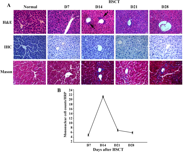
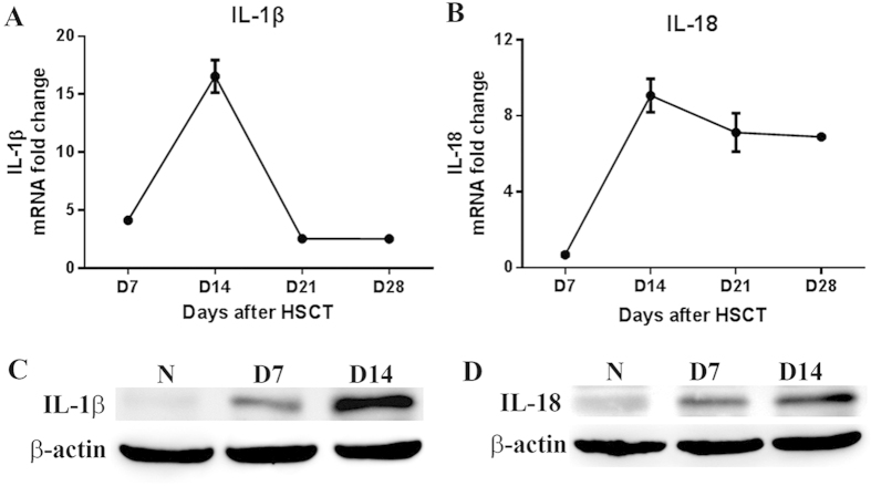
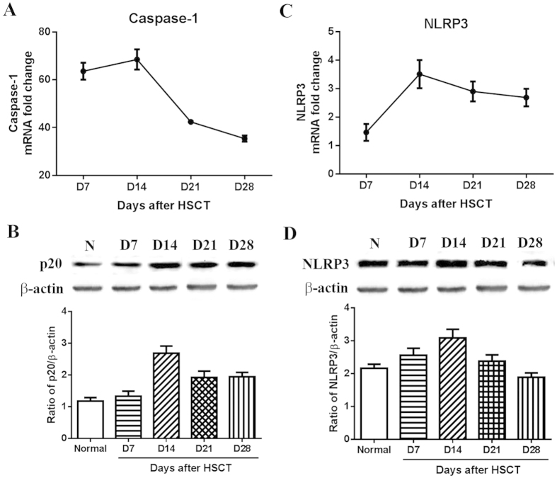
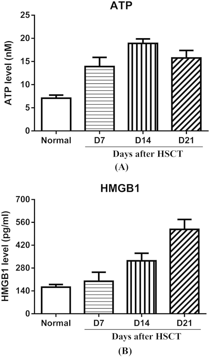
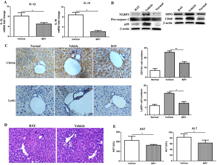
The aim of this study was to evaluate the role of NLRP3 inflammasome on BU/CY-induced liver inflammation in mice after HSCT. HSCT mice model was established through infusion of 5 × 10(6) bone marrow mononuclear cells after conditioned with BU/CY. On day 7, 14, 21 and 28 after HSCT, mice were sacrificed for analysis of liver inflammation, cytokine secretion, NLRP3 expression and caspase-1 activation as well as release of ATP and high-mobility group protein B1 (HMGB1). Furthermore, NLRP3 selective inhibitor (BAY 11-7082) was administrated into mice after HSCT to evaluate its effects on liver inflammation. Severe liver inflammation and damage with elevated secretion of IL-1β and IL-18 were found in mice after HSCT. Meanwhile, elevated expressions of NLRP3 and caspase-1 activation in liver were found. In addition, increased release of ATP and HMGB1 were observed. Selective inhibition of NLRP3 decreased caspase-1 activation and secretion of IL-1β and IL-18. Furthermore, NLRP3 inhibition also reduced infiltration of macrophages and neutrophils and improved liver function. In conclusion, NLRP3 was involved in BU/CY-induced liver inflammation after HSCT and selectively inhibited it ameliorated liver inflammation and improved liver function, suggesting targeting NLRP3 might be a new approach in the prophylaxis of liver inflammation after HSCT.
Figures
Similar articles
-
[The relationship between NLRP3 inflammsomes expression and liver damage after allogeneic hematopoietic stem cell transplantation in mice model].Zhonghua Xue Ye Xue Za Zhi. 2014 Aug;35(8):684-8. doi: 10.3760/cma.j.issn.0253-2727.2014.08.003. Zhonghua Xue Ye Xue Za Zhi. 2014. PMID: 25152112 Chinese.
-
Chenodeoxycholic acid activates NLRP3 inflammasome and contributes to cholestatic liver fibrosis.Oncotarget. 2016 Dec 20;7(51):83951-83963. doi: 10.18632/oncotarget.13796. Oncotarget. 2016. PMID: 27924062 Free PMC article.
-
Tripartite-motif protein 30 negatively regulates NLRP3 inflammasome activation by modulating reactive oxygen species production.J Immunol. 2010 Dec 15;185(12):7699-705. doi: 10.4049/jimmunol.1001099. Epub 2010 Nov 3. J Immunol. 2010. PMID: 21048113
-
Regulation and Function of the Nucleotide Binding Domain Leucine-Rich Repeat-Containing Receptor, Pyrin Domain-Containing-3 Inflammasome in Lung Disease.Am J Respir Cell Mol Biol. 2016 Feb;54(2):151-60. doi: 10.1165/rcmb.2015-0231TR. Am J Respir Cell Mol Biol. 2016. PMID: 26418144 Free PMC article. Review.
-
Extracellular Adenosine Triphosphate (eATP) and Its Metabolite, Extracellular Adenosine (eAdo), as Opposing "Yin-Yang" Regulators of Nlrp3 Inflammasome in the Trafficking of Hematopoietic Stem/Progenitor Cells.Front Immunol. 2021 Jan 29;11:603942. doi: 10.3389/fimmu.2020.603942. eCollection 2020. Front Immunol. 2021. PMID: 33584673 Free PMC article. Review.
Cited by
-
NLRP3 Inflammasome: A Promising Therapeutic Target for Drug-Induced Toxicity.Front Cell Dev Biol. 2021 Apr 12;9:634607. doi: 10.3389/fcell.2021.634607. eCollection 2021. Front Cell Dev Biol. 2021. PMID: 33912556 Free PMC article. Review.
-
NLRP3 regulates platelet integrin αIIbβ3 outside-in signaling, hemostasis and arterial thrombosis.Haematologica. 2018 Sep;103(9):1568-1576. doi: 10.3324/haematol.2018.191700. Epub 2018 May 24. Haematologica. 2018. PMID: 29794149 Free PMC article.
-
NRF2 -617 C/A Polymorphism Impacts Proinflammatory Cytokine Levels, Survival, and Transplant-Related Mortality After Hematopoietic Stem Cell Transplantation in Adult Patients Receiving Busulfan-Based Conditioning Regimens.Front Pharmacol. 2020 Dec 15;11:563321. doi: 10.3389/fphar.2020.563321. eCollection 2020. Front Pharmacol. 2020. PMID: 33384597 Free PMC article.
-
Metformin pretreatment ameliorates busulfan-induced liver endothelial toxicity during haematopoietic stem cell transplantation.PLoS One. 2023 Oct 26;18(10):e0293311. doi: 10.1371/journal.pone.0293311. eCollection 2023. PLoS One. 2023. PMID: 37883349 Free PMC article.
-
Inflammasomes-New Contributors to Blood Diseases.Int J Mol Sci. 2022 Jul 23;23(15):8129. doi: 10.3390/ijms23158129. Int J Mol Sci. 2022. PMID: 35897704 Free PMC article. Review.
References
-
- Copelan E. A. Hematopoietic stem-cell transplantation. N Engl J Med 354, 1813–1826 (2006). - PubMed
-
- Pidala J. Progress in hematopoietic cell transplantation. Cancer Control 18, 212–213 (2011). - PubMed
-
- Ho V. T., Linden E., Revta C. & Richardson P. G. Hepatic veno-occlusive disease after hematopoietic stem cell transplantation: review and update on the use of defibrotide. Semin Thromb Hemost 33, 373–388 (2007). - PubMed
-
- Pidala J. Graft-vs-host disease following allogeneic hematopoietic cell transplantation. Cancer Control 18, 268–276 (2011). - PubMed
Publication types
MeSH terms
Substances
LinkOut - more resources
Full Text Sources
Other Literature Sources
Medical
Miscellaneous
