. 2016 Feb 12;291(7):3124-34.
doi: 10.1074/jbc.M115.703710. Epub 2015 Dec 3.

Interaction between a Domain of the Negative Regulator of the Ras-ERK Pathway, SPRED1 Protein, and the GTPase-activating Protein-related Domain of Neurofibromin Is Implicated in Legius Syndrome and Neurofibromatosis Type 1

Affiliations

Interaction between a Domain of the Negative Regulator of the Ras-ERK Pathway, SPRED1 Protein, and the GTPase-activating Protein-related Domain of Neurofibromin Is Implicated in Legius Syndrome and Neurofibromatosis Type 1

Yasuko Hirata et al. J Biol Chem. .

Abstract

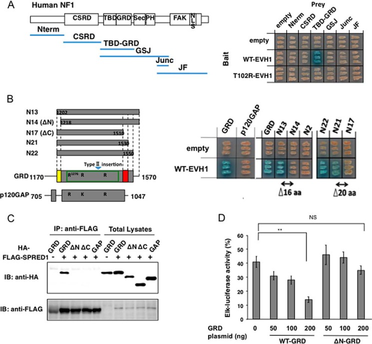
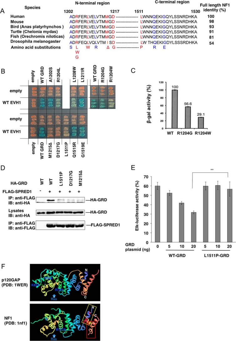
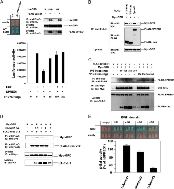
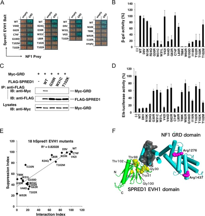
Constitutional heterozygous loss-of-function mutations in the SPRED1 gene cause a phenotype known as Legius syndrome, which consists of symptoms of multiple café-au-lait macules, axillary freckling, learning disabilities, and macrocephaly. Legius syndrome resembles a mild neurofibromatosis type 1 (NF1) phenotype. It has been demonstrated that SPRED1 functions as a negative regulator of the Ras-ERK pathway and interacts with neurofibromin, the NF1 gene product. However, the molecular details of this interaction and the effects of the mutations identified in Legius syndrome and NF1 on this interaction have not yet been investigated. In this study, using a yeast two-hybrid system and an immunoprecipitation assay in HEK293 cells, we found that the SPRED1 EVH1 domain interacts with the N-terminal 16 amino acids and the C-terminal 20 amino acids of the GTPase-activating protein (GAP)-related domain (GRD) of neurofibromin, which form two crossing α-helix coils outside the GAP domain. These regions have been shown to be dispensable for GAP activity and are not present in p120(GAP). Several mutations in these N- and C-terminal regions of the GRD in NF1 patients and pathogenic missense mutations in the EVH1 domain of SPRED1 in Legius syndrome reduced the binding affinity between the EVH1 domain and the GRD. EVH1 domain mutations with reduced binding to the GRD also disrupted the ERK suppression activity of SPRED1. These data clearly demonstrate that SPRED1 inhibits the Ras-ERK pathway by recruiting neurofibromin to Ras through the EVH1-GRD interaction, and this study also provides molecular basis for the pathogenic mutations of NF1 and Legius syndrome.

Keywords: GTPase-activating protein (GAP); Ras protein; human genetics; mitogen-activated protein kinase (MAPK); negative regulation; protein domain; signal transduction.


Figures

FIGURE 1.
FIGURE 1.
Yeast two-hybrid assay to detect EVH1 and neurofibromin interacting domains. A, yeast strains carrying pGBKT7-hEVH1(1–136) and the indicated neurofibromin domains cloned in pGADT7 were restreaked on a filter paper and stained for an in situ galactosidase assay. B, interaction between the EVH1 domain and deletion mutants of the GRD. Left, schematic view of the structural comparison between the NF-1 GRD and the GAP domain of p120GAP. The green box in the GRD indicates that it is minimum domain with full GTPase-activating activity (36). R and K in the GRD and GAP indicate conserved residues for Ras-GTP binding. Right, cDNAs of p120-RasGAP(705–1047) or neurofibromin GRD deletion mutants were subcloned into pACT2. N13 means full-length GRD(1170–1570). Yeasts were restreaked on a filter paper and stained for an in situ galactosidase assay. aa, amino acid. C, binding of EVH1 and GRD mutants in HEK293 cells. HEK293 cells grown in 10-cm dishes were transiently transfected with HA-tagged deletion mutants of GRD plasmids (1.5 μg/dish) together with FLAG-tagged SPRED1 expression plasmid (1 μg/dish). The immunoprecipitates (IP) with anti-FLAG antibody were immunoblotted (IB) with anti-HA and anti-FLAG antibodies. D, enhancement of SPRED1-dependent ERK suppression by GRD. HEK293 cells grown in 12-well plates were transfected with Elk-1 reporter plasmid, hSPRED1 expression vector (10 ng/well), together with indicated amounts of WT-GRD or ΔN-GRD expression vectors. After 24 h, cells were treated with 50 ng/ml EGF for 6 h, and luciferase activity was then measured. The Elk-luciferase activity of cells with EGF stimulation without transfecting SPRED1 and GRD plasmids is standardized as 100%. NS, not significant; **, p < 0.01.
FIGURE 2.
FIGURE 2.
GRD mutations in the N-terminal extended region reduce EVH1 binding affinity. A, amino acid sequences of the N- and C-terminal EVH1 binding region of GRDs from various species. Red letters indicate amino acids whose mutations severely affected binding to the EVH1 domain. Blue letters show amino acids whose mutations had little effect on the binding. B, in situ yeast two-hybrid assay of the interaction between EVH1 and NF1 GRD mutants. Yeast strains carrying pGBKT7-hEVH1 and the indicated GRD mutants in pGADT7 were restreaked on a filter paper and stained for an in situ galactosidase assay. C, quantitative β-gal assay of yeast strains carrying pGBKT7-hEVH1 and pGADT7-GRD with indicated mutations. D, binding of EVH1 and GRD mutants in HEK293 cells. HEK293 cells were transiently transfected with HA-tagged GRD mutants and FLAG-tagged SPRED1. The immunoprecipitates (IP) with anti-FLAG antibody were immunoblotted (IB) with anti-HA and anti-FLAG antibodies. E, GRDL1511P showed a weaker effect on SPRED1-mediated ERK suppression than WT-GRD. HEK293 cells were transfected with Elk-1 reporter plasmid and hSPRED1 expression vector, together with indicated amounts of WT-GRD or GRDL1511P expression vectors. After 24 h, cells were treated with 50 ng/ml EGF for 6 h and luciferase activity was then measured. The Elk-luciferase activity of cells with EGF stimulation without transfecting SPRED1 and GRD plasmids is standardized as 100%. F, ribbon drawing structures of GRDs of p120GAP and NF1. Blue, N-terminal region. **, p < 0.01. Beige color, C-terminal region. The positions of Leu1511, Leu1208, Met1212, and Asp1217 are shown.
FIGURE 3.
FIGURE 3.
GRD-EVH1 interaction is independent of the GRD-Ras interaction. A, effect of the GAP-null mutant of GRD on SPRED1 suppression activity. Upper panels, in situ yeast two-hybrid assay and IP-Western blotting assay for the interaction between WT EVH1 and R1276P mutant GRD. Lower panel, reversal of the effect of hSPRED1 on R1276P GRD. HEK293 cells were transfected with or without 10 ng of hSPRED1 cDNA in the presence of increased concentrations of R1276P GRD cDNA. EGF-induced Elk-1 reporter activity was measured 1 day after transfection. IP, immunoprecipitation; IB, immunoblot. B, comparison of the binding of GRD between SPRED1 and Ras. HEK293 cells were transfected with FLAG-tagged SPRED1 (2 μg), WT (0.5 μg), or V12-H-Ras (0.25 μg) and Myc-GRD (2 μg) expression plasmids. Cell extracts were immunoprecipitated with anti-FLAG antibody and then blotted with anti-FLAG or anti-Myc antibodies. C, effect of the overexpression of Ras on the SPRED1-GRD interaction. HEK293 cells were transfected with FLAG-SPRED1 cDNA (3 μg) and Myc-GRD cDNA (0.1 μg) together with increased concentrations of FLAG-tagged WT or V12 H-Ras cDNA plasmids. One day after transfection, cell extracts were immunoprecipitated with anti-Myc antibody and then blotted with anti-Myc or anti-FLAG antibodies. D, effect of the overexpression of the SPRED1-EVH1 domain on the V12-HRas-GRD interaction. HEK293 cells were transfected with 500 ng of FLAG-Ras cDNA and 1.5 μg of Myc-GRD cDNA together with increased concentrations of HA-tagged SPRED1 (EVH1) cDNA plasmids. One day after transfection, cell extracts were immunoprecipitated with anti-FLAG antibody and then blotted with anti-Myc or anti-FLAG antibodies. E, yeast strains carrying pGBKT7-EVH1s from mSpred1–3 (mS1, mS2, and mS3) and pGADT7-GRD were restreaked on a filter paper and stained for an in situ galactosidase assay (upper panels). A quantitative β-galactosidase (β-gal) assay was performed and normalized against the GRD-hSPRED1 EVH1 interaction.
FIGURE 4.
FIGURE 4.
Effects of EVH1 mutations on the EVH1-GRD interaction and ERK suppression activity. A and B, yeast strains carrying pGBKT7-hEVH1 with indicated mutations and pGADT7-GRD were restreaked on a filter paper and stained for an in situ galactosidase assay (A) and a quantitative β-gal assay (B). Error bars denote the mean ± S.D. of triplicate experiments. C, lack of interaction of the GRD with mutant EVH1 domains in 293 cells. Cells were transfected with Myc-tagged GRD and indicated FLAG-tagged SPRED1 mutants. Immunoprecipitates (IP) with anti-FLAG antibody were blotted with anti-Myc antibody. IB, immunoblot. D, ERK suppression activity by mutant SPREDs. HEK293 cells were transfected with the Elk-1 reporter system and 30 ng of mutant SPRED1 cDNAs. One day after transfection, HEK293 cells were stimulated with 50 ng/ml EGF for 6 h, and luciferase activity was then measured. E, correlation between ERK suppression activity and GRD binding activity of mutant EVH1 domains. Plotting of the EVH1-GRD interaction index and ERK suppression activity index is shown. The EVH1-GRD interaction index is calculated from B. WT EVH1 β-gal activity is standardized as 100%. ERK suppression index (calculated from D) = ((Elk-1 reporter with mutant Spred1) − (Elk-1 reporter with WT Spred1))/(Elk-1 reporter without Spred1). F, in silico modeling of the EVH1 domain (green) and GRD (light blue). The side chains of the arginine residues in charge of binding to Ras (Arg1276 and Arg1416) in GRD are shown in magenta. The N- and C-terminal helices in the GRD, which are important for EVH1 binding, are covered with a gray surface. Plausible residues in EVH1 for interaction with GRD suggested from the results of yeast two-hybrid assay and ERK suppression assay (Gly30, Trp31, Gly62, Thr88, Gly100, and Thr102) are shown in yellow.
FIGURE 5.
FIGURE 5.
Information of the family carrying R24Q mutation. Upper panel, pedigree of the family. Open symbols indicate unaffected individuals; lined symbols indicate affected individuals. *, p < 0.05. Table, clinical phenotypes of individuals carrying R24Q mutation. CALM, café-au-lait spot; −, not present; u, unknown.

References

    1. Yoshimura A. (2009) Regulation of cytokine signaling by the SOCS and Spred family proteins. Keio J. Med. 58, 73–83 - PubMed
    1. Bundschu K., Walter U., and Schuh K. (2007) Getting a first clue about SPRED functions. BioEssays 29, 897–907 - PubMed
    1. Kato R., Nonami A., Taketomi T., Wakioka T., Kuroiwa A., Matsuda Y., and Yoshimura A. (2003) Molecular cloning of mammalian Spred-3 which suppresses tyrosine kinase-mediated Erk activation. Biochem. Biophys. Res. Commun. 302, 767–772 - PubMed
    1. Nonami A., Taketomi T., Kimura A., Saeki K., Takaki H., Sanada T., Taniguchi K., Harada M., Kato R., and Yoshimura A. (2005) The Sprouty-related protein, Spred-1, localizes in a lipid raft/caveola and inhibits ERK activation in collaboration with caveolin-1. Genes Cells 10, 887–895 - PubMed
    1. Impagnatiello M. A., Weitzer S., Gannon G., Compagni A., Cotten M., and Christofori G. (2001) Mammalian sprouty-1 and -2 are membrane-anchored phosphoprotein inhibitors of growth factor signaling in endothelial cells. J. Cell Biol. 152, 1087–1098 - PMC - PubMed

Publication types

MeSH terms

Substances

Supplementary concepts