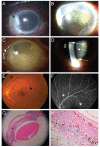
Sympathetic Ophthalmia after Ocular Wasp Sting
- PMID: 26635462
- PMCID: PMC4668261
- DOI: 10.3341/kjo.2015.29.6.435
Sympathetic Ophthalmia after Ocular Wasp Sting
Conflict of interest statement
Figures

Similar articles
-
Management of Corneal Bee Sting Injuries.Semin Ophthalmol. 2017;32(2):177-181. doi: 10.3109/08820538.2015.1045301. Semin Ophthalmol. 2017. PMID: 26161915
-
Wasp sting of the cornea: a case treated with amniotic membrane transplantation.Graefes Arch Clin Exp Ophthalmol. 2013 Mar;251(3):1039-40. doi: 10.1007/s00417-012-2072-y. Epub 2012 Jun 9. Graefes Arch Clin Exp Ophthalmol. 2013. PMID: 22678716 No abstract available.
-
[Sympathetic ophthalmia, case report].Oftalmologia. 2006;50(3):48-51. Oftalmologia. 2006. PMID: 17144506 Romanian.
-
Sympathetic ophthalmia.Int Ophthalmol Clin. 2002 Summer;42(3):179-85. doi: 10.1097/00004397-200207000-00019. Int Ophthalmol Clin. 2002. PMID: 12131594 Review. No abstract available.
-
Corneal Emergencies.Top Companion Anim Med. 2015 Sep;30(3):74-80. doi: 10.1053/j.tcam.2015.07.006. Epub 2015 Jul 9. Top Companion Anim Med. 2015. PMID: 26494498 Review.
Cited by
-
Ocular Injury Caused by the Sprayed Venom of the Asian Giant Hornet (Vespa mandarinia).Case Rep Ophthalmol. 2020 Aug 6;11(2):430-435. doi: 10.1159/000508911. eCollection 2020 May-Aug. Case Rep Ophthalmol. 2020. PMID: 32999672 Free PMC article.
-
An Atypical Presentation of Sympathetic Ophthalmia in an Intact Globe Following Mechanical Fall: A Case Report and Literature Review.Vision (Basel). 2021 Feb 21;5(1):11. doi: 10.3390/vision5010011. Vision (Basel). 2021. PMID: 33669961 Free PMC article.
References
Publication types
MeSH terms
Substances
LinkOut - more resources
Full Text Sources
Other Literature Sources
Medical
Research Materials

