Review
. 2015 Nov 17:8:67.
doi: 10.3389/fnmol.2015.00067. eCollection 2015.

Protein and Signaling Networks in Vertebrate Photoreceptor Cells

Affiliations
Review

Protein and Signaling Networks in Vertebrate Photoreceptor Cells

Karl-Wilhelm Koch et al. Front Mol Neurosci. .

Abstract

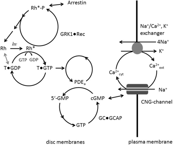
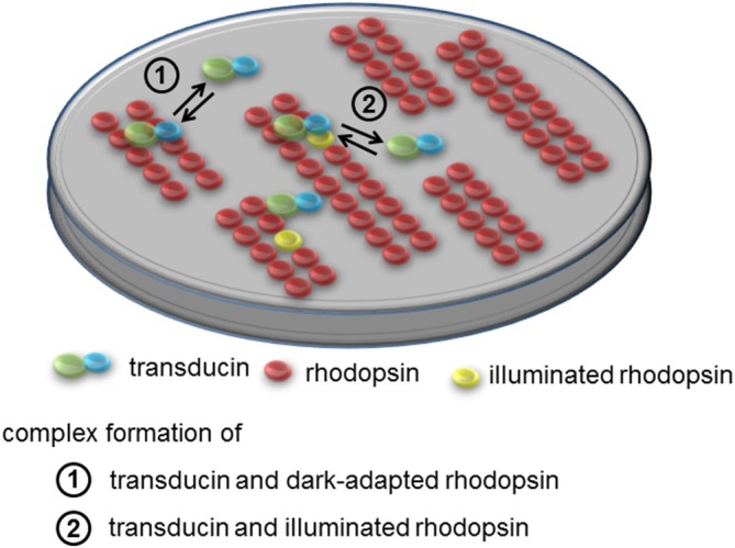
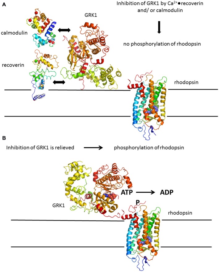
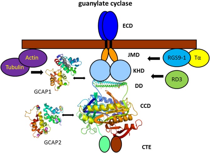
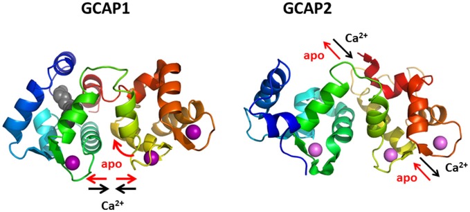
Vertebrate photoreceptor cells are exquisite light detectors operating under very dim and bright illumination. The photoexcitation and adaptation machinery in photoreceptor cells consists of protein complexes that can form highly ordered supramolecular structures and control the homeostasis and mutual dependence of the secondary messengers cyclic guanosine monophosphate (cGMP) and Ca(2+). The visual pigment in rod photoreceptors, the G protein-coupled receptor rhodopsin is organized in tracks of dimers thereby providing a signaling platform for the dynamic scaffolding of the G protein transducin. Illuminated rhodopsin is turned off by phosphorylation catalyzed by rhodopsin kinase (GRK1) under control of Ca(2+)-recoverin. The GRK1 protein complex partly assembles in lipid raft structures, where shutting off rhodopsin seems to be more effective. Re-synthesis of cGMP is another crucial step in the recovery of the photoresponse after illumination. It is catalyzed by membrane bound sensory guanylate cyclases (GCs) and is regulated by specific neuronal Ca(2+)-sensor proteins called guanylate cyclase-activating proteins (GCAPs). At least one GC (ROS-GC1) was shown to be part of a multiprotein complex having strong interactions with the cytoskeleton and being controlled in a multimodal Ca(2+)-dependent fashion. The final target of the cGMP signaling cascade is a cyclic nucleotide-gated (CNG) channel that is a hetero-oligomeric protein located in the plasma membrane and interacting with accessory proteins in highly organized microdomains. We summarize results and interpretations of findings related to the inhomogeneous organization of signaling units in photoreceptor outer segments.

Keywords: cGMP; calcium-binding proteins; multi-protein complexes; phototransduction; second messenger signaling.


Figures

Figure 1
Figure 1
Main signaling steps in phototransduction. Photo-activation of rhodopsin (Rh to Rh*) leads to GDP/GTP exchange at the G protein transducin (T) which in turn activates its effector PDE. Hydrolysis of cGMP is catalyzed by activated PDE; resynthesis of cGMP by guanylate cyclase (GC) is under control of a negative Ca2+-feedback involving the GC-activating proteins (GCAP) Ca2+-sensor proteins. Ca2+ enters the cell via the cyclic nucleotide-gated (CNG)-channel and is extruded by the exchanger. Rh* is phosphorylated by GRK1, when inhibition by Ca2+-bound recoverin is relieved. Arrestin can bind to phosphorylated Rh* preventing further activation of transducin.
Figure 2
Figure 2
Supramolecular organization of rhodopsin and interaction with transducin. Rhodopsin is present in tracks of dimers in the disc membrane. In the dark rhodopsin-transducin complexes form with submicromolar affinity that is characterized by very fast association and dissociation rates. Movements of transducin can be described as dynamic hopping on rhodopsin supramolecular assemblies thus constituting “dynamic scaffolding”. Apparent dissociation rates of transducin from dark-adapted rhodopsin are >300-fold faster than corresponding rates from light-activated rhodopsin.
Figure 3
Figure 3
Rhodopsin phosphorylation by GRK1. (A) Deactivation of rhodopsin is under control of a Ca2+-feedback loop involving Ca2+-sensor proteins recoverin and calmodulin. Both Ca2+-binding proteins have non-overlapping binding sites in GRK1 and act in a synergetic mode, for example by increasing the Ca2+-sensitivity of GRK1 regulation. (B) Inhibition of GRK1 is relieved at decreasing Ca2+-concentration after illumination leading to phosphorylation of rhodopsin. Structures were prepared with pymol; corresponding PDB codes are: 1F88 for rhodopsin (Palczewski et al., 2000); 3C51 for GRK1 (Singh et al., 2008); 1JSA for recoverin (Ames et al., 1997); 1CDM for calmodulin (Meador et al., 1993).
Figure 4
Figure 4
Vertebrate photoreceptor GC and interacting proteins. Photoreceptor GCs contain several domains denoted as extracellular domain (ECD, which in rod outer segment is present in the intradiscal lumen; TM, transmembrane domain; JMD, juxtamembrane domain; KHD, kinase homology domain; DD, dimerization domain; CCD, cyclase catalytic domain; and CTE, a C-terminal extension). The tertiary structure of the DD and CCD is adapted from the solved three-dimensional structure of soluble GCs (Ma et al., ; Allerston et al., 2013) that display a high sequence homology with membrane bound GCs in these domains (PDB codes: 3HLS and 3UVJ). Assembly and topography of GC domains, in particular of the DD and CCD is arbitrarily chosen. GCAP1 and GCAP2 activate the target GC at low cytoplasmic Ca2+-concentration bringing the cell back to the dark state. The exact regions of interaction and/or regulation by GCAPs are a matter of debate (see main text). Interaction with other proteins was shown by biochemical procedures, but physiological meaning is lacking so far. Structures of GCAP1 and GCAP2 are based on the published x-ray and NMR-structure, respectively (GCAP1: 2R2I, Stephen et al., ; GCAP2: 1JBA, Ames et al., 1999).
Figure 5
Figure 5
Protein dynamics of GCAP1 and GCAP2. Binding and dissociation of Ca2+ (violet spheres) triggers different conformational changes and movements of secondary structural elements, a twisted accordion-like movement in GCAP1 and a piston-like movement of one α-helix (yellow) in GCAP2.

References

    1. Allerston C. K., von Delft F., Gileadi O. (2013). Crystal structures of the catalytic domain of human soluble guanylate cyclase. PLoS One 8:e57644. 10.3410/f.718029832.793480138 - DOI - PMC - PubMed
    1. Ames J. B., Dizhoor A. M., Ikura M., Palczewski K., Stryer L. (1999). Three-dimensional structure of guanylyl cyclase activating protein-2, a calcium-sensitive modulator of photoreceptor guanylyl cyclases. J. Biol. Chem. 274, 19329–19337. 10.1074/jbc.274.27.19329 - DOI - PubMed
    1. Ames J. B., Ishima R., Tanaka T., Gordon J. I., Stryer L., Ikura M. (1997). Molecular mechanics of calcium-myristoyl switches. Nature 389, 198–202. - PubMed
    1. Ames J. B., Levay K., Wingard J. N., Lusin J. D., Slepak V. Z. (2006). Structural basis for calcium-induced inhibition of rhodopsin kinase by recoverin. J. Biol. Chem. 281, 37237–37245. 10.1074/jbc.m606913200 - DOI - PubMed
    1. Arshavsky V. Y., Burns M. E. (2012). Photoreceptor signaling: supporting vision across a wide range of light intensities. J. Biol. Chem. 287, 1620–1626. 10.1074/jbc.r111.305243 - DOI - PMC - PubMed

LinkOut - more resources