. 2015 Nov 23:9:453.
doi: 10.3389/fncel.2015.00453. eCollection 2015.

Dual Nitrergic/Cholinergic Control of Short-Term Plasticity of Corticostriatal Inputs to Striatal Projection Neurons

Affiliations

Dual Nitrergic/Cholinergic Control of Short-Term Plasticity of Corticostriatal Inputs to Striatal Projection Neurons

Craig P Blomeley et al. Front Cell Neurosci. .

Abstract

The ability of nitric oxide and acetylcholine to modulate the short-term plasticity of corticostriatal inputs was investigated using current-clamp recordings in BAC mouse brain slices. Glutamatergic responses were evoked by stimulation of corpus callosum in D1 and D2 dopamine receptor-expressing medium spiny neurons (D1-MSNs and D2-MSN, respectively). Paired-pulse stimulation (50 ms intervals) evoked depressing or facilitating responses in subgroups of both D1-MSNs and D2 MSNs. In both neuronal types, glutamatergic responses of cells that displayed paired-pulse depression were not significantly affected by the nitric oxide donor S-nitroso-N-acetylpenicillamine (SNAP; 100 μM). Conversely, in D1-MSNs and D2-MSNs that displayed paired-pulse facilitation, SNAP did not affect the first evoked response, but significantly reduced the amplitude of the second evoked EPSP, converting paired-pulse facilitation into paired-pulse depression. SNAP also strongly excited cholinergic interneurons and increased their cortical glutamatergic responses acting through a presynaptic mechanism. The effects of SNAP on glutamatergic response of D1-MSNs and D2-MSN were mediated by acetylcholine. The broad-spectrum muscarinic receptor antagonist atropine (25 μM) did not affect paired-pulse ratios and did not prevent the effects of SNAP. Conversely, the broad-spectrum nicotinic receptor antagonist tubocurarine (10 μM) fully mimicked and occluded the effects of SNAP. We concluded that phasic acetylcholine release mediates feedforward facilitation in MSNs through activation of nicotinic receptors on glutamatergic terminals and that nitric oxide, while increasing cholinergic interneurons' firing, functionally impairs their ability to modulate glutamatergic inputs of MSNs. These results show that nitrergic and cholinergic transmission control the short-term plasticity of glutamatergic inputs in the striatum and reveal a novel cellular mechanism underlying paired-pulse facilitation in this area.

Keywords: cholinergic interneuron; microcircuit; nitrergic interneuron; nitric oxide; striatum.


Figures
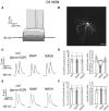
Figure 1
Figure 1
Paired pulse facilitation in D1-MSNs is converted into paired-pulse depression by SNAP. (A) A D1-MSN displayed typical electrophysiological properties when injected with current steps (20 pA increments). Rheobase current was 220 pA. (B) Neurobiotin staining of the neuron in A revealed dense dendritic arborizations and dendritic spines. Scale bar = 50 μm. (C) In a D1-MSNs, paired-pulse facilitation (at 50 ms interval) was observed in control solution (which contained GABAA and GABAB receptor antagonists), but was converted into paired-pulse depression by SNAP, that reduced the amplitude of the second evoked EPSP. These effects were reversed after SNAP washout. Each trace in this and subsequent figures showing paired pulse responses is the average of at least 50 consecutive responses. (D) Effects of SNAP on the first evoked response and the paired-pulse ratio of D1-MSNs that displayed paired-pulse facilitation in control solution. In this and in the following figures, asterisks indicate that the effects of certain treatment were statistical significant (*p < 0.05 and ***p < 0.001) in every cell tested. (E) In another D1-MSN, paired-pulse depression of corticostriatal responses was present in control solution. In this case, application of SNAP failed to significantly affect these responses. (F) Effects of SNAP on the first evoked response and the paired-pulse ratio of D1-MSNs that displayed paired-pulse depression in control solution.
Figure 2
Figure 2
SNAP converted paired-pulse facilitation into paired-pulse depression in D2-MSNs. (A) Responses of a D2-MSN to current injections (20 pA increments). Rheobase current was 80 pA. (B) Neurobiotin staining of the neuron in A revealed its dendritic arborization and spines. Scale bar = 50 μm. (C) In a D2-MSNs paired-pulse facilitation was present in control solution (which contained GABAA and GABAB receptor antagonists), but was reversibly converted into paired-pulse depression by SNAP, that reduced the amplitude of the second evoked EPSP. Each trace in this and other figures showing paired pulse responses is the average of 50 consecutive responses. (D) Effects of SNAP on the first evoked response and the paired-pulse ratio of D2-MSNs that displayed paired-pulse facilitation in control solution. (E) In another D2-MSN, paired-pulse depression of corticostriatal responses was present in control solution. In this case, application of SNAP failed to significantly affect these responses. (F) Effects of SNAP on the first evoked response and on paired-pulse ratio of D2-MSNs that displayed paired-pulse depression in control solution.
Figure 3
Figure 3
SNAP excites cholinergic interneurons and facilitates their glutamatergic inputs. (A) Bath application of SNAP caused a strong increase in spontaneous firing frequency in a cholinergic interneuron recorded in cell-attached configuration. (B) Average frequency of spontaneous spikes in cholinergic interneurons recorded in cell-attached or whole-cell configurations in control solution and in the presence of SNAP. A significant (p < 0.05) increase was observed in each interneuron tested. (C) SNAP increased the glutamatergic responses evoked in voltage-clamp by paired-pulse stimulation of corticostriatal fibers (each trace is the average of 100 consecutive responses). (D) Effects of SNAP on the amplitude of the first response and the paired-pulse ratio in cholinergic interneurons. (E) Effects of SNAP on miniature EPSCs in a cholinergic interneuron. (F) Average effects of SNAP on the cumulative probability of amplitude and inter-event interval distribution of spontaneous miniature EPSCs. The average amplitude and frequency of mini EPSCs were both significantly larger in the presence of SNAP.
Figure 4
Figure 4
Blocking nicotinic receptors mimics and occludes the effects of SNAP on paired-pulse facilitation in D1 and D2-MSNs. (A) In a D2-MSN, paired-pulse facilitation was converted into paired-pulse depression by SNAP. After washout of the SNAP effects, application of atropine failed to affect the evoked responses. In the presence of atropine, SNAP had similar effects as when applied in control solution. Each trace is the average of 50 consecutive responses. (B) Average effects on paired-pulse ratio of SNAP, atropine and combined SNAP plus atropine application in a population of facilitating cells comprising four D1-MSNs and eight D2-MSNs. In each neuron tested, the paired-pulse ratios were significantly lower in SNAP and in atropine plus SNAP than in control. (C) In another D2-MSN, paired-pulse facilitation was converted into paired-pulse depression by SNAP. After washout of the SNAP effects, application of tubocurarine also converted paired-pulse facilitation into paired-pulse depression. When applied in the presence of tubocurarine, SNAP failed to affect the evoked responses. (D) Effects on paired-pulse ratio of SNAP, tubocurarine and SNAP plus tubocurarine in a population of facilitating MSNs comprising four D1-MSNs and eight D2-MSNs. In each neuron tested, the paired-pulse ratios was significantly lower in the presence of SNAP alone, tubocurarine alone or tubocurarine plus SNAP than in control solution. (E) Bath-application of nicotine, that induces profound desensitization of nicotinic receptors, converted paired-pulse facilitation into paired-pulse depression in a D1-MSNs (left) and a D2-MSN (right).
Figure 5
Figure 5
Microcircuit underlying short term plasticity of MSN responses. Diagram of the microcircuit proposed to be involved in the short-term plasticity of corticostriatal inputs to facilitating MSNs. Cortical inputs impinge on MSNs and on cholinergic interneurons (CI). In the absence of nitric oxide (top), ACh is released after the first stimulus and activates nicotinic receptors on the cortical terminals impinging on MSNs. As a result, the second pulse causes a larger release of glutamate from these terminals (paired-pulse facilitation). When nitric oxide is released by nitrergic interneurons (bottom), cholinergic interneurons are excited, but presynaptic nicotinic receptors are no longer activated, possibly as a result of vesicle depletion or receptor desensitization. As a result, the second pulse releases less glutamate than the first (paired-pulse depression).

References

    1. Beatty J. A., Sullivan M. A., Morikawa H., Wilson C. J. (2012). Complex autonomous firing patterns of striatal low-threshold spike interneurons. J. Neurophysiol. 108, 771–781. 10.1152/jn.00283.2012 - DOI - PMC - PubMed
    1. Bennett B. D., Callaway J. C., Wilson C. J. (2000). Intrinsic membrane properties underlying spontaneous tonic firing in neostriatal cholinergic interneurons. J. Neurosci. 20, 8493–8503. 10.1016/0306-4522(87)90021-2 - DOI - PMC - PubMed
    1. Blomeley C., Bracci E. (2005). Excitatory effects of serotonin on rat striatal cholinergic interneurones. J. Physiol. 569, 715–721. 10.1113/jphysiol.2005.098269 - DOI - PMC - PubMed
    1. Centonze D., Pisani A., Bonsi P., Giacomini P., Bernardi G., Calabresi P. (2001). Stimulation of nitric oxide-cGMP pathway excites striatal cholinergic interneurons via protein kinase G activation. J. Neurosci. 21, 1393–1400. - PMC - PubMed
    1. Ding J. B., Guzman J. N., Peterson J. D., Goldberg J. A., Surmeier D. J. (2010). Thalamic gating of corticostriatal signaling by cholinergic interneurons. Neuron 67, 294–307. 10.1016/j.neuron.2010.06.017 - DOI - PMC - PubMed