Review
. 2015 Nov 24:6:283.
doi: 10.3389/fphar.2015.00283. eCollection 2015.

Role of ErbB Receptors in Cancer Cell Migration and Invasion

Affiliations
Review

Role of ErbB Receptors in Cancer Cell Migration and Invasion

Aline Appert-Collin et al. Front Pharmacol. .

Abstract

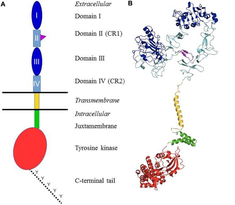
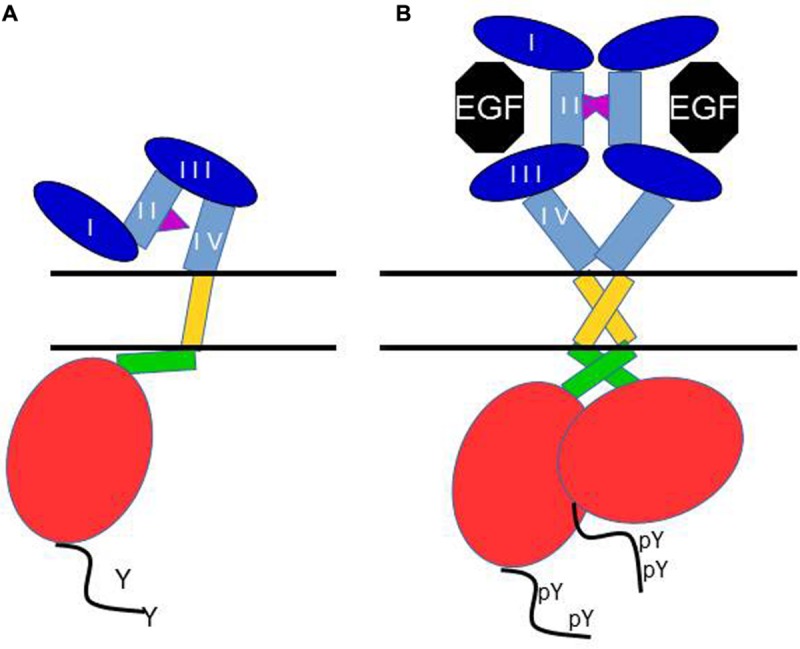
Growth factors mediate their diverse biologic responses (regulation of cellular proliferation, differentiation, migration and survival) by binding to and activating cell-surface receptors with intrinsic protein kinase activity named receptor tyrosine kinases (RTKs). About 60 RTKs have been identified and can be classified into more than 16 different receptor families. Their activity is normally tightly controlled and regulated. Overexpression of RTK proteins or functional alterations caused by mutations in the corresponding genes or abnormal stimulation by autocrine growth factor loops contribute to constitutive RTK signaling, resulting in alterations in the physiological activities of cells. The ErbB receptor family of RTKs comprises four distinct receptors: the EGFR (also known as ErbB1/HER1), ErbB2 (neu, HER2), ErbB3 (HER3) and ErbB4 (HER4). ErbB family members are often overexpressed, amplified, or mutated in many forms of cancer, making them important therapeutic targets. EGFR has been found to be amplified in gliomas and non-small-cell lung carcinoma while ErbB2 amplifications are seen in breast, ovarian, bladder, non-small-cell lung carcinoma, as well as several other tumor types. Several data have shown that ErbB receptor family and its downstream pathway regulate epithelial-mesenchymal transition, migration, and tumor invasion by modulating extracellular matrix (ECM) components. Recent findings indicate that ECM components such as matrikines bind specifically to EGF receptor and promote cell invasion. In this review, we will present an in-depth overview of the structure, mechanisms, cell signaling, and functions of ErbB family receptors in cell adhesion and migration. Furthermore, we will describe in a last part the new strategies developed in anti-cancer therapy to inhibit ErbB family receptor activation.

Keywords: ErbB receptors; cancer; cell signaling; epithelial-mesenchymal transition; migration.


Figures

FIGURE 1
FIGURE 1
Structural organization of the ErbB/HER receptors. (A) Shows a schematic representation of the different domains. The extracellular part of the receptors is composed of four domains: I and III = ligand-binding domain (dark blue); II and IV = cysteine-rich domains (light blue). The domain II contains the dimerization arm (purple). It is followed by the single transmembrane domain (yellow), a juxtamembrane domain (green), the tyrosine kinase domain (red) and a C-terminal tail which contains the main tyrosines that are phosphorylated upon receptor activation (dotted line). (B) Presents the structures of different domains of the human EGFR which have been established through X-ray crystallography or solution RMN. Color coding is identical to (A). Protein Data Bank (PDB) accession codes are as follows: ectodomain (in closed unliganded conformation), 3QWQ, transmembrane region, 2KS1, juxtamembrane domain, 1Z9I; kinase domain, 3W32. No structure is available for the C-terminal tail.
FIGURE 2
FIGURE 2
Schematic of the current view of main structural events in the activation of the EGF receptor. In (A) the receptor is depicted in its monomeric, unliganded, inactive form. The dimerization arm of the extracellular domain II binds to domain IV, and the juxtamembrane domain interacts with membrane phospholipids. In (B) binding of EGF to one monomer to domains I and III induces a conformation change which makes the dimerization arm available for interaction with another extended ligand-bound monomer, and causes dimerization. This conformational change is accompanied by the formation of an anti-parallel interaction between the two juxtamembrane domains, and thus an asymmetric “head to tail” interaction of the two kinase domains, resulting in allosteric activation of the kinase, and C-terminal tail tyrosine phosphorylation.

References

    1. Alaoui-Jamali M. A., Morand G. B., da Silva S. D. (2015). ErbB polymorphisms: insights and implications for response to targeted cancer therapeutics. Front. Genet. 6:17 10.3389/fgene.2015.00017 - DOI - PMC - PubMed
    1. Al Moustafa A. E., Achkhar A., Yasmeen A. (2012). EGF-receptor signaling and epithelial-mesenchymal transition in human carcinomas. Front. Biosci. (Schol. Ed.) 4:671–684. 10.2741/S292 - DOI - PubMed
    1. Arpel A., Sawma P., Spenle C., Fritz J., Meyer L., Garnier N., et al. (2014). Transmembrane domain targeting peptide antagonizing ErbB2/Neu inhibits breast tumor growth and metastasis. Cell Rep. 8 1714–1721. 10.1016/j.celrep.2014.07.044 - DOI - PubMed
    1. Bedard P. L., Cardoso F., Piccart-Gebhart M. J. (2009). Stemming resistance to HER-2 targeted therapy. J. Mammary Gland Biol. Neoplasia 14 55–66. 10.1007/s10911-009-9116-x - DOI - PubMed
    1. Bennasroune A., Fickova M., Gardin A., Dirrig-Grosch S., Aunis D., Cremel G., et al. (2004a). Transmembrane peptides as inhibitors of ErbB receptor signaling. Mol. Biol. Cell 15 3464–3474. 10.1091/mbc.E03-10-0753 - DOI - PMC - PubMed