Review
. 2015 Nov 25:6:347.
doi: 10.3389/fphys.2015.00347. eCollection 2015.

The Measurement of Reversible Redox Dependent Post-translational Modifications and Their Regulation of Mitochondrial and Skeletal Muscle Function

Affiliations
Review

The Measurement of Reversible Redox Dependent Post-translational Modifications and Their Regulation of Mitochondrial and Skeletal Muscle Function

Philip A Kramer et al. Front Physiol. .

Abstract

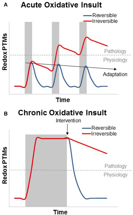
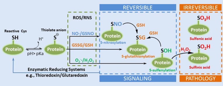
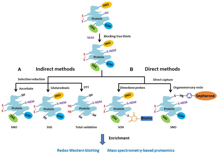
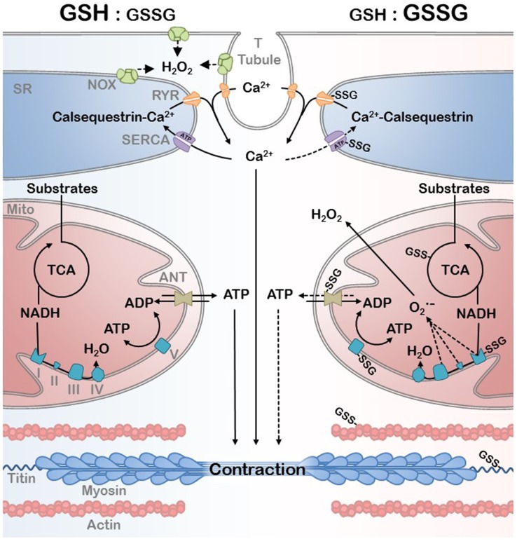
Mitochondrial oxidative stress is a common feature of skeletal myopathies across multiple conditions; however, the mechanism by which it contributes to skeletal muscle dysfunction remains controversial. Oxidative damage to proteins, lipids, and DNA has received the most attention, yet an important role for reversible redox post-translational modifications (PTMs) in pathophysiology is emerging. The possibility that these PTMs can exert dynamic control of muscle function implicates them as a mechanism contributing to skeletal muscle dysfunction in chronic disease. Herein, we discuss the significance of thiol-based redox dependent modifications to mitochondrial, myofibrillar, and excitation-contraction (EC) coupling proteins with an emphasis on how these changes could alter skeletal muscle performance under chronically stressed conditions. A major barrier to a better mechanistic understanding of the role of reversible redox PTMs in muscle function is the technical challenges associated with accurately measuring the changes of site-specific redox PTMs. Here we will critically review current approaches with an emphasis on sample preparation artifacts, quantitation, and specificity. Despite these challenges, the ability to accurately quantify reversible redox PTMs is critical to understanding the mechanisms by which mitochondrial oxidative stress contributes to skeletal muscle dysfunction in chronic diseases.

Keywords: glutathionylation; mitochondria; myofibrils; post-translational modification; redox signaling; skeletal muscle.


Figures

Figure 1
Figure 1
Theoretical graph of the transient and additive nature of reversible and irreversible redox PTMs in the context of skeletal muscle physiology and disease. (A) Acute oxidative insults (shaded area) can cause a swift and transient increase in reversible oxidative PTMs. Irreversible PTMs rely on protein degradation and turnover, while most reversible modifications can be cleared by the GSH/GSSG and Trx(red)/Trx(ox) redox couples. Subsequent acute oxidative insults can be deleterious if the modifications become irreversible, but this is not the case for reversible modifications, which, can cause transcriptional activation of the antioxidant response (adaptation, preconditioning, or hormesis) to suppress PTM formation. (B) Chronic oxidative insults can result in an oxidized redox status which persists throughout the duration of the stress. As with aging, many metabolic and functional skeletal muscle defects that result from this oxidized redox status can be rapidly reversed by restoring a more reduced redox environment. In contrast, irreversible oxidative modifications, however, can persist after the end of the oxidative insult.
Figure 2
Figure 2
A simplified diagram of the formation of thiol redox PTMs. A reactive Cys can exist as thiolate anion under physiological pH. The thioate anion is reactive with different ROS or RNS to form several types of reversible redox PTMs such as SNO, SSG, and SOH. SOH can be further oxidized into irreversible sulfinic and sulfonic acid (Adapted from Filomeni et al. Cell Death and Differentiation 2005, Filomeni et al., 2005).
Figure 3
Figure 3
Schematic of the general approaches for measuring different reversible thiol-based redox PTMs. The free thiols are typically blocked by alkylation with NEM as the initial step. (A) In the indirect methods, the different types of modified cysteines are selectively reduced to free thiols by using individual sets of reagents. (B) In the direct methods, dimendone probe is often used to react with SOH, and organomercury resin for SNO. The reduced or labeled protein samples are then subjected to enrichment and measurement by either gel-based or MS-based proteomic approaches.
Figure 4
Figure 4
EC coupling and bioenergetics network in a reduced (high GSH:GSSG ratio) and oxidative (low GSH:GSSG ratio) redox environment. Ca2+ handling by the SR and the ATP generated by the mitochondria are the primary substrates of myofibril contraction. Oxidative PTM can affect the mitochondrial efficiency as well as EC coupling during redox signaling and the oxidative stress associated with disease. Protein S-Glutathioylation of critical mitochondrial, SR, and myofibrillar proteins are depicted as well as the primary sources of H2O2.

References

    1. Adlam V. J., Harrison J. C., Porteous C. M., James A. M., Smith R. A., Murphy M. P., et al. . (2005). Targeting an antioxidant to mitochondria decreases cardiac ischemia-reperfusion injury. FASEB J. 19, 1088–1095. 10.1096/fj.05-3718com - DOI - PubMed
    1. Alegre-Cebollada J., Kosuri P., Giganti D., Eckels E., Rivas-Pardo J. A., Hamdani N., et al. . (2014). S-glutathionylation of cryptic cysteines enhances titin elasticity by blocking protein folding. Cell 156, 1235–1246. 10.1016/j.cell.2014.01.056 - DOI - PMC - PubMed
    1. Alleman R. J., Katunga L. A., Nelson M. A., Brown D. A., Anderson E. J. (2014). The “Goldilocks Zone” from a redox perspective-Adaptive vs. deleterious responses to oxidative stress in striated muscle. Front. Physiol. 5:358. 10.3389/fphys.2014.00358 - DOI - PMC - PubMed
    1. Anderson E. J., Lustig M. E., Boyle K. E., Woodlief T. L., Kane D. A., Lin C. T., et al. . (2009). Mitochondrial H2O2 emission and cellular redox state link excess fat intake to insulin resistance in both rodents and humans. J. Clin. Invest. 119, 573–581. 10.1172/JCI37048 - DOI - PMC - PubMed
    1. Anderson E. J., Neufer P. D. (2006). Type II skeletal myofibers possess unique properties that potentiate mitochondrial H(2)O(2) generation. Am. J. Physiol. Cell Physiol. 290, C844–C851. 10.1152/ajpcell.00402.2005 - DOI - PubMed

LinkOut - more resources