Review
. 2015 Nov 26:6:1805.
doi: 10.3389/fpsyg.2015.01805. eCollection 2015.

Prefrontal Cortex and Social Cognition in Mouse and Man

Affiliations
Review

Prefrontal Cortex and Social Cognition in Mouse and Man

Lucy K Bicks et al. Front Psychol. .

Abstract

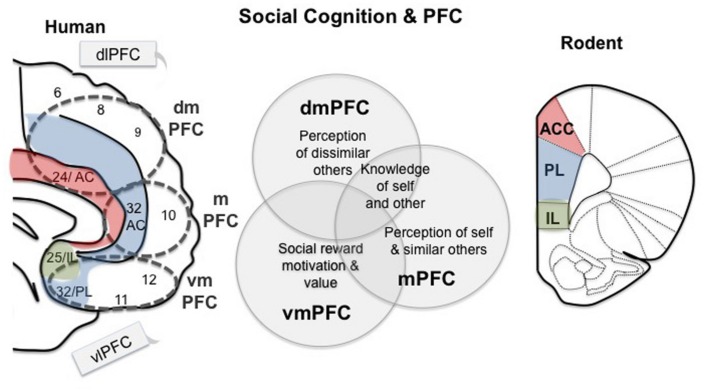
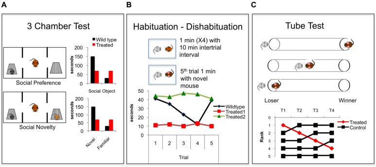
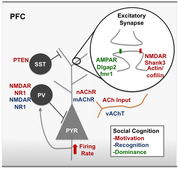
Social cognition is a complex process that requires the integration of a wide variety of behaviors, including salience, reward-seeking, motivation, knowledge of self and others, and flexibly adjusting behavior in social groups. Not surprisingly, social cognition represents a sensitive domain commonly disrupted in the pathology of a variety of psychiatric disorders including Autism Spectrum Disorder (ASD) and Schizophrenia (SCZ). Here, we discuss convergent research from animal models to human disease that implicates the prefrontal cortex (PFC) as a key regulator in social cognition, suggesting that disruptions in prefrontal microcircuitry play an essential role in the pathophysiology of psychiatric disorders with shared social deficits. We take a translational perspective of social cognition, and review three key behaviors that are essential to normal social processing in rodents and humans, including social motivation, social recognition, and dominance hierarchy. A shared prefrontal circuitry may underlie these behaviors. Social cognition deficits in animal models of neurodevelopmental disorders like ASD and SCZ have been linked to an altered balance of excitation and inhibition (E/I ratio) within the cortex generally, and PFC specifically. A clear picture of the mechanisms by which altered E/I ratio in the PFC might lead to disruptions of social cognition across a variety of behaviors is not well understood. Future studies should explore how disrupted developmental trajectory of prefrontal microcircuitry could lead to altered E/I balance and subsequent deficits in the social domain.

Keywords: autism; prefrontal cortex; schizophrenia; social behavior; social cognition.


Figures

FIGURE 1
FIGURE 1
Working model for prefrontal regions involved in social cognition in human and mouse. Medial regions of the prefrontal cortex (PFC) are specifically related to social behavior, while the lateral regions, dlPFC and vlPFC, are sometimes active during social tasks, but are considered ‘domain general.’ The dmPFC is involved in perceptions of others as well as cooperation (Amodio and Frith, 2006; Mitchell et al., 2006). The mPFC has also been associated with perceptions of others, but some research suggests that it is more strongly associated with perceptions of self and similar others (Johnson et al., 2002; Mitchell et al., 2006; Mitchell, 2009). Ventral regions of the PFC are involved in social reward and punishment, motivation and ‘value’ (including economic) (de Quervain et al., 2004; Fehr and Camerer, 2007; Kohls et al., 2012). Parts of these divisions in the human brain share homology with the rodent PFC, as indicated. VmPFC contains BA 25, which is homologous to the rodent IL region, and area 32 is homologous to the PL. Area 24 in humans shares homology with the rodent ACC. These regions thus may play a shared role in social cognition across mammalian lineages.
FIGURE 2
FIGURE 2
Common behavioral paradigms for studying social cognition in rodents. (A) The three chamber test (Moy et al., 2004). In the first phase social preference is assessed. A focal mouse chooses between a social target and an object and time spent investigating both is measured and compared. In the second phase social novelty preference is assessed when a novel mouse is added and the focal mouse chooses to investigate a novel vs. familiar mouse. Graphs show common findings demonstrating the natural wildtype (black bars) propensity to investigate a social target more than an object, and to investigate a novel mouse more than a familiar mouse. Red bars demonstrate a hypothetical treated group showing no social preference and no novel social preference. (B) The Habituation – Dishabituation paradigm (Thor and Holloway, 1982) in which a juvenile mouse is presented to a focal mouse, usually in the home cage, for four consecutive 1 min trials with an intertrial interval of 10 min. A novel juvenile is presented on the fifth trial. The graph shows commonly reported wildtype social investigation time (black), which decreases over the four trials and then increases with the presentation of the novel mouse on the fifth trial, demonstrating recognition of a novel animal. Hypothetical red data shows floor levels of social investigation, similar to that seen when animals are treated with NMDAR antagonists (Zou et al., 2008; Jeevakumar et al., 2015). Data indicated by the green line shows a ceiling level of social investigation showing hypothetical intact social motivation and decreased social recognition. This effect is seen in animals lacking the oxytocin gene (Ferguson et al., 2000). (C) The tube test. Tests for dominance by placing two mice into a tube and recording which mouse forces the other to back out of the tube (Lindzey et al., 1966). A fictitious experiment is shown in which the rank of the four control mice (black) is compared over time. The top ranked mouse is treated (red) and drops rank within the hierarchy. This effect is similar to that seen when the synaptic efficacy within the PFC is decreased (Wang et al., 2011a).
FIGURE 3
FIGURE 3
Modulators of social cognition in the rodent PFC. Red text represents nodes of the circuit that, when disrupted, decrease social motivation. For example, synaptic scaffolding proteins on excitatory synapses like Shank3 and IRSp53 have been associated with social motivation in the PFC, as have cytoskeleton remodelers, actin and cofilin. NMDARs at excitatory synapses are also a key node of the social motivation circuit. ACh input to the PFC and nicotinic receptors have also been shown to modulate social motivation, however, it is unclear which cell types are important for ACh action or whether these effects are pre or post-synaptic. Blue text represents nodes of the circuit that, when disrupted, decrease social recognition. For example, disrupting gabaergic neurotransmission by removing the NR1 subunit on cortical gabaergic interneurons disrupts social recognition. Green text represents nodes of the circuit that are involved in dominance behavior. For example, bidirectional modulation of AMPARs and mutations in the fmr1 gene. ACh, acetylcholine; AMPAR, α-amino-3-hydroxy-5-methyl-4-isoxazolepropionic acid receptor; dlgap2, disks large-associated protein 2, fmr1, fragile X mental retardation 1; IRSp53, insulin receptor substrate protein of 53 kDa, mAchR, muscarinic acetylcholine receptor, nAChR, nicotinic acetylcholine receptor, NMDAR, N-Methyl-D-aspartate receptor, PV, parvalbumin postitive interneuron, vAChT, vesicular acetylcholine transporter. See text for references.

References

    1. Adolphs R. (2009). The social brain: neural basis of social knowledge. Annu. Rev. Psychol. 60 693–716. 10.1146/annurev.psych.60.110707.163514 - DOI - PMC - PubMed
    1. Akbarian S., Kim J. J., Potkin S. G., Hagman J. O., Tafazzoli A., Bunney W. E., Jr., et al. (1995). Gene expression for glutamic acid decarboxylase is reduced without loss of neurons in prefrontal cortex of schizophrenics. Arch. Gen. Psychiatry 52 258–266. 10.1001/archpsyc.1995.03950160008002 - DOI - PubMed
    1. Amir R. E., Van Den Veyver I. B., Wan M., Tran C. Q., Francke U., Zoghbi H. Y. (1999). Rett syndrome is caused by mutations in X-linked MECP2, encoding methyl-CpG-binding protein 2. Nat. Genet. 23 185–188. 10.1038/13810 - DOI - PubMed
    1. Amodio D. M., Frith C. D. (2006). Meeting of minds: the medial frontal cortex and social cognition. Nat. Rev. Neurosci. 7 268–277. 10.1038/nrn1884 - DOI - PubMed
    1. Anderson P. K. (1961). Density, social structure, and nonsocial environment in house-mouse populations and the implications for regulation of numbers∗. Transact. N. Y. Acad. Sci. 23 447–451. 10.1111/j.2164-0947.1961.tb01373.x - DOI - PubMed

LinkOut - more resources