. 2015 Nov 10:6:1256.
doi: 10.3389/fmicb.2015.01256. eCollection 2015.

Niemeyer Virus: A New Mimivirus Group A Isolate Harboring a Set of Duplicated Aminoacyl-tRNA Synthetase Genes

Affiliations

Niemeyer Virus: A New Mimivirus Group A Isolate Harboring a Set of Duplicated Aminoacyl-tRNA Synthetase Genes

Paulo V M Boratto et al. Front Microbiol. .

Abstract

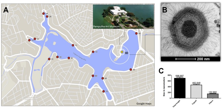
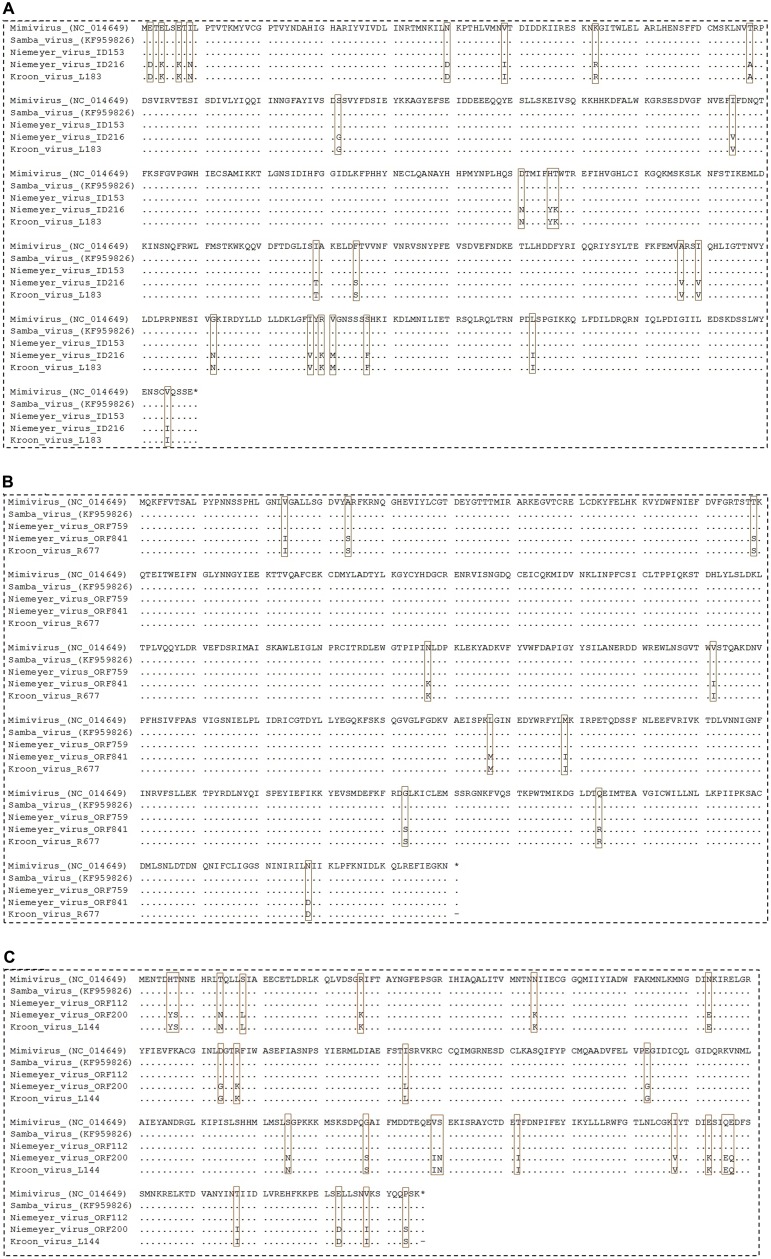
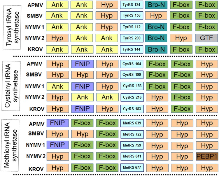
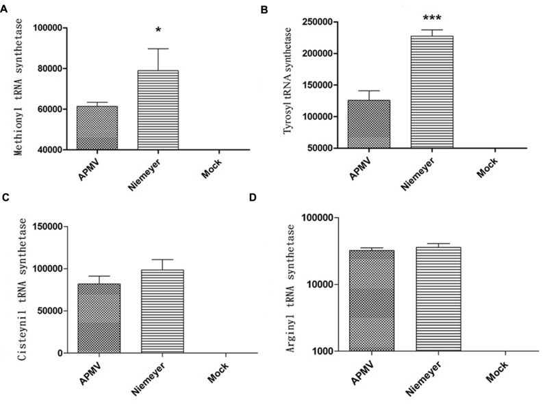
It is well recognized that gene duplication/acquisition is a key factor for molecular evolution, being directly related to the emergence of new genetic variants. The importance of such phenomena can also be expanded to the viral world, with impacts on viral fitness and environmental adaptations. In this work we describe the isolation and characterization of Niemeyer virus, a new mimivirus isolate obtained from water samples of an urban lake in Brazil. Genomic data showed that Niemeyer harbors duplicated copies of three of its four aminoacyl-tRNA synthetase genes (cysteinyl, methionyl, and tyrosyl RS). Gene expression analysis showed that such duplications allowed significantly increased expression of methionyl and tyrosyl aaRS mRNA by Niemeyer in comparison to APMV. Remarkably, phylogenetic data revealed that Niemeyer duplicated gene pairs are different, each one clustering with a different group of mimivirus strains. Taken together, our results raise new questions about the origins and selective pressures involving events of aaRS gain and loss among mimiviruses.

Keywords: Mimiviridae; Niemeyer virus; aminoacyl-tRNA synthetase; gene duplication; giant virus isolation.


Figures

FIGURE 1
FIGURE 1
Niemeyer virus (NYMV) collection site, morphometric analysis, and electron microscopy. (A) Shows a map of the Pampulha Lagoon, from where the samples were collected. Red dots: collection points; yellow dot: NYMV isolation site. (B) Shows an image of a NYMV particle, observed in electron microscopy. (C) Shows the size, in nanometers, of different components of the NYMV particle: only the fibers, only the capsid and the particle as a whole.
FIGURE 2
FIGURE 2
Alignment of NYMV aaRs sequences. (A) Cysteinyl-tRNA synthetase, (B) methionyl-tRNA synthetase, and (C) tyrosyl-tRNA synthetase sequences. In this analysis sequences from Acanthamoeba polyphaga mimivirus (APMV), the prototype of Mimivirus, and sequences from Samba virus (SMBV) and Kroon virus (KROV) were used. Brown boxes highlight polymorphisms in the alignments.
FIGURE 3
FIGURE 3
Neighboring gene synteny analyses of aminoacyl-tRNA synthetase. Three upstream and downstream neighbor genes of each aaRS predicted in the genome of NYMV and other mimivirus were checked. Ank, ankyrin repeat family protein; Hyp, Hypothetical Protein; F-box, F-box protein family; Bro-n, bro-n family protein; GTF, glycosyltransferase; FNIP, FNIP repeat-containing protein; PEBP1, phosphatidylethanolamine-binding protein. NYMV 1 and 2 refer to each copy of the aaRS genes.
FIGURE 4
FIGURE 4
Aminoacyl-tRNA-synthetase (aaRS) mRNA expression by NYMV. (A) Methionyl tRNA synthetase, (B) tyrosyl tRNA synthetase, (C) cysteinyl tRNA synthetase, and (D) arginyl tRNA synthetase. Relative gene expression analyses were performed using the ΔΔCt method and normalized to the expression of 18S ribosomal RNA [18S rDNA) and the viral RNA helicase mRNA (and calibrated with the lower value (=1)]. The values were subjected in different combinations to one-way ANOVA tests and Bonferroni post-tests (95% confidence intervals). Differences between groups were considered significant when the p-values were smaller than 0.05 (asterisks). Y axis represents relative abundance.
FIGURE 5
FIGURE 5
(A) One step growth curves of NYMV and APMV in Acanthamoeba castellanii. At 6 hpi cells are similar negative control cell (data not shown). (B) A. castellanii cells counting at different time points (0, 8, and 24 hpi with APMV or NYMV). (C) Images obtained from cells of A. castellanii infected with NYMV and APMV at different time points after infection.
FIGURE 6
FIGURE 6
Phylogenetic reconstruction of mimiviruses including the Brazilian Niemeyer mimivirus strain, based on the Polymerase-B amino acid sequences, using MEGA5 software (Maximum-likelihood – 1,000 bootstrap replicates). The NYMV (highlighted by black dot) clustered with other Brazilian mimivirus isolates (triangles), as well as other members of mimivirus lineage A. For each sequence, the gene identification number is indicated.
FIGURE 7
FIGURE 7
Phylogenetic analysis based on sequences of (A) Arginyl-tRNA synthetase, (B) Methionyl-tRNA synthetase, (C) Tyrosyl-tRNA synthetase, and (D) Cysteinyl-tRNA synthetase predicted in the genome of NYMV, and orthologous sequences obtained from the NCBI database. The trees were inferred by using MEGA5 software (Neighbor-joining – 1,000 bootstrap replicates). The aaRS encoded by NYMV are highlighted by black dots. For each sequence, the gene identification number is indicated. Red and blue boxes indicates the different copies of NYMV aaRS.

References

    1. Abrahão J. S., Boratto P. V. M., Dornas F. P., Silva L. C., Campos R. K., Almeida G. M., et al. (2014). Growing a giant: evaluation of the virological parameters for mimivirus production. J. Virol. Methods 207 6–11. 10.1016/j.jviromet.2014.06.001 - DOI - PubMed
    1. Arslan D., Legendre M., Seltzer V., Abergel C., Claverie J. M. (2011). Distant Mimivirus relative with a larger genome highlights the fundamental features of Megaviridae. Proc. Natl. Acad. Sci. U.S.A. 108 17486–17491. 10.1073/pnas.1110889108 - DOI - PMC - PubMed
    1. Boughalmi M., Saadi H., Pagnier I., Colson P., Fournous G., Raoult D., et al. (2013). High-throughput isolation of giant viruses of the Mimiviridae and Marseilleviridae families in the Tunisian environment. Environ. Microbiol. 15 2000–2007. 10.1111/1462-2920.12068 - DOI - PubMed
    1. Campos R. K., Boratto P. V., Assis F. L., Aguiar E. R., Silva L. C., Albarnaz J. D., et al. (2014). Samba virus: a novel mimivirus from a giant rain forest, the Brazilian Amazon. Virol. J. 11 95 10.1186/1743-422X-11-95 - DOI - PMC - PubMed
    1. Dornas F. P., Silva L. C., de Almeida G. M., Campos R. K., Boratto P. V., Franco-Luiz A. P., et al. (2014). Acanthamoeba polyphaga mimivirus stability in environmental and clinical substrates: implications for virus detection and isolation. PLoS ONE 9:e87811 10.1371/journal.pone.0087811 - DOI - PMC - PubMed

LinkOut - more resources