. 2016 Apr;68(4):944-52.
doi: 10.1002/art.39499.

Lupus Nephritis IgG Induction of Calcium/Calmodulin-Dependent Protein Kinase IV Expression in Podocytes and Alteration of Their Function

Affiliations

Lupus Nephritis IgG Induction of Calcium/Calmodulin-Dependent Protein Kinase IV Expression in Podocytes and Alteration of Their Function

Kunihiro Ichinose et al. Arthritis Rheumatol. 2016 Apr.

Abstract

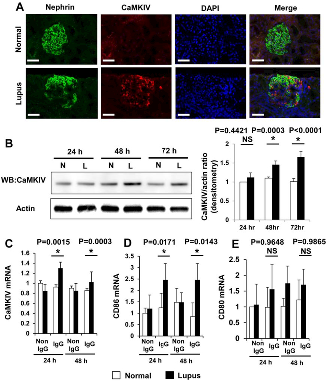
Objective: Kidney podocytes and their slit diaphragms prevent urinary protein loss. T cells from patients with systemic lupus erythematosus display increased expression of calcium/calmodulin-dependent protein kinase IV (CaMKIV). The present study was undertaken to investigate the role of CaMKIV in podocyte function in lupus nephritis (LN).

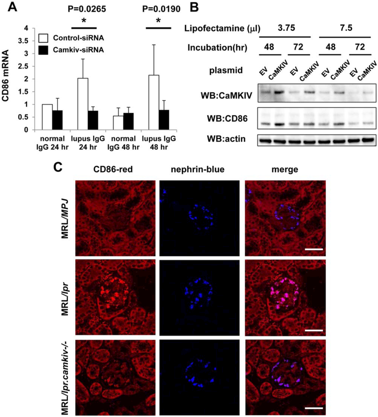
Methods: We treated kidney podocytes with IgG derived from healthy individuals or patients with LN and then analyzed gene expression using a DNA microarray. The localization of IgG in podocytes was analyzed by immunofluorescence staining, with or without silencing of neonatal Fc receptor (FcRn). In addition, we silenced CAMK4 in podocytes and analyzed the expression of selected genes. We also examined the expression of CD86 in kidney podocytes from MRL/lpr, MRL/lpr.camkiv(-/-), and MRL/MPJ mice by in situ hybridization.

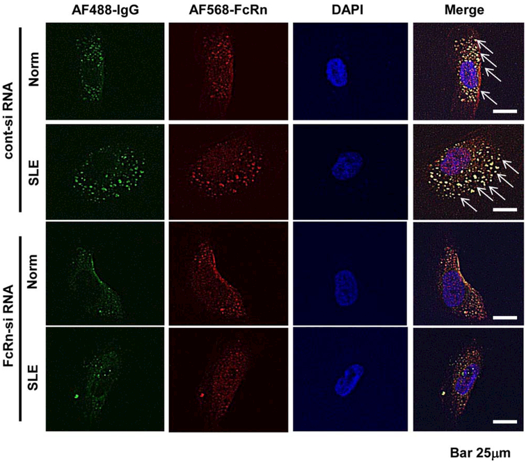
Results: We found that exposure of podocytes to IgG resulted in entry of IgG into the cytoplasm. IgG entered podocytes via the FcRn because less IgG was found in the cytoplasm of podocytes treated with FcRn small interfering RNA. DNA microarray studies of podocytes exposed to LN-derived IgG revealed up-regulation of genes related to the activation of immune cells or podocyte damage. Interestingly, CD86 expression decreased after silencing CAMK4 in podocytes. Also, in situ hybridization experiments showed that the expression of CD86 was reduced in podocytes from MRL/lpr.camkiv(-/-) mice.

Conclusion: LN-derived IgG enters podocytes and up-regulates CAMK4, which is followed by increased expression of genes known to be linked to podocyte damage and T cell activation. Targeted inhibition of CAMK4 in podocytes may prove to be clinically useful in patients with LN.


Figures

Figure 1.
Figure 1.
A, Electrophoresis of whole serum (WS) or fractionated serum IgG derived from a systemic lupus erythematosus (SLE) patient (L) and a normal subject (N). Molecular weight marker (M) is shown at the right. B, Scatterplot of entire gene set considered by microarray analysis to be expressed in podocytes (P < 0.05). The position of each dot on the scatterplot corresponds to the normalized average signal intensity (log scale) of a single gene. The normalized average signal intensity after exposure to SLE IgG and normal IgG is shown on the x and y axes, respectively. Broken red lines represent normal IgG:SLE IgG ratios of 2.0 (top; 2-fold greater expression in normal IgG) and 0.5 (bottom; 2-fold greater expression in SLE IgG). Black broken line is the mean.
Figure 2.
Figure 2.
IgG entry into podocytes via the neonatal Fc receptor (FcRn). Localization of IgG derived from a normal subject (Norm) and a patient with systemic lupus erythematosus (SLE) nephritis (Alexa Fluor [AF] 488 stained) in podocytes was analyzed by immunofluorescence staining, with or without silencing of FcRn (Alexa Fluor 568 stained), the receptor of IgG, using FcRn small interfering RNA (siRNA) or control siRNA. Arrows indicate entry of IgG into the cytoplasm, which did not occur when FcRn was silenced.
Figure 3.
Figure 3.
A, Coexpression of nephrin and calcium/calmodulin-dependent protein kinase IV (CaMKIV) in the glomeruli of normal subjects and patients with lupus nephritis. Bars = 50 μm. B–E, Lupus serum induction of the expression of CaMKIV protein (B), CaMKIV mRNA (C), CD86 mRNA (D), and CD80 mRNA (E) after incubation for the indicated times. Podocytes (cell line) were stimulated with IgG derived from sera obtained from normal controls (N) and lupus nephritis patients (L). Protein expression of CaMKIV was determined by Western blotting (WB) (B). Actin was included as a loading control. The Western blotting results were quantified by densitometry and are shown at the right. Values are the mean ± SD of 4–8 subjects per group. * = P < 0.05 versus normal controls. NS = not significant.
Figure 4.
Figure 4.
A, Suppression of CD86 expression by silencing of calcium/calmodulin-dependent protein kinase IV (CaMKIV) in human podocytes exposed to IgG derived from normal subjects and from patients with lupus nephritis. Human podocytes were exposed to IgG for 24 or 48 hours and were then transfected with CAMK4 or control scrambled small interfering RNA (siRNA). Values are the mean ± SD of 4 subjects per group. * = P < 0.05 versus control siRNA. B, Expression of mRNA for CD86 in human podocytes following forced expression of CAMK4. Cultured human podocytes were transfected with either empty vector (EV) or pCMV-sport6 CaMKIV. After 48 or 72 hours, cell lysates were collected, and the expression of CaMKIV, CD86, and actin (loading control) protein was examined by Western blotting (WB). C, Reduced expression of CD86 in the glomeruli of lupus-prone mice lacking CaMKIV. The coexpression of CD86 mRNA and nephrin mRNA in the glomeruli of MRL/MpJ, MRL/lpr, and MRL/lpr.camkiv−/− mice was analyzed by in situ hybridization. Bars = 50 μm.

Similar articles

Cited by

References

    1. Tsokos GC. Systemic lupus erythematosus. N Engl J Med 2011; 365:2110–21. - PubMed
    1. Greka A, Mundel P. Cell biology and pathology of podocytes. Annu Rev Physiol 2012;74:299–323. - PMC - PubMed
    1. Kraft SW, Schwartz MM, Korbet SM, Lewis EJ. Glomerular podocytopathy in patients with systemic lupus erythematosus. J Am Soc Nephrol 2005;16:175–9. - PubMed
    1. Trivedi S, Zeier M, Reiser J. Role of podocytes in lupus nephritis. Nephrol Dial Transplant 2009;24:3607–12. - PubMed
    1. Juang YT, Wang Y, Solomou EE, Li Y, Mawrin C, Tenbrock K, et al. Systemic lupus erythematosus serum IgG increases CREM binding to the IL-2 promoter and suppresses IL-2 production through CaMKIV. J Clin Invest 2005;115:996–1005. - PMC - PubMed

Publication types

MeSH terms