. 2015 Dec 4;10(12):e0144378.
doi: 10.1371/journal.pone.0144378. eCollection 2015.

Stochastic Mesocortical Dynamics and Robustness of Working Memory during Delay-Period

Affiliations

Stochastic Mesocortical Dynamics and Robustness of Working Memory during Delay-Period

Melissa Reneaux et al. PLoS One. .

Erratum in

Abstract

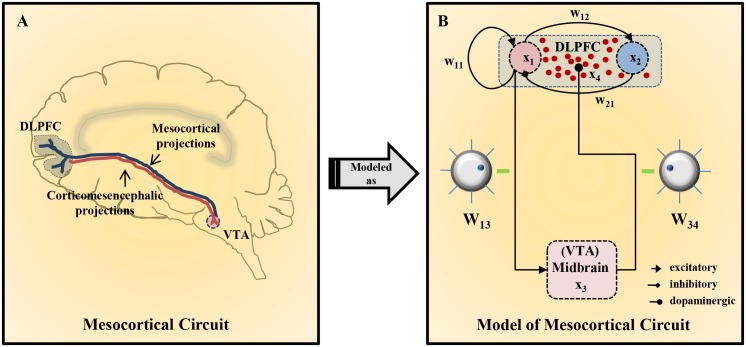
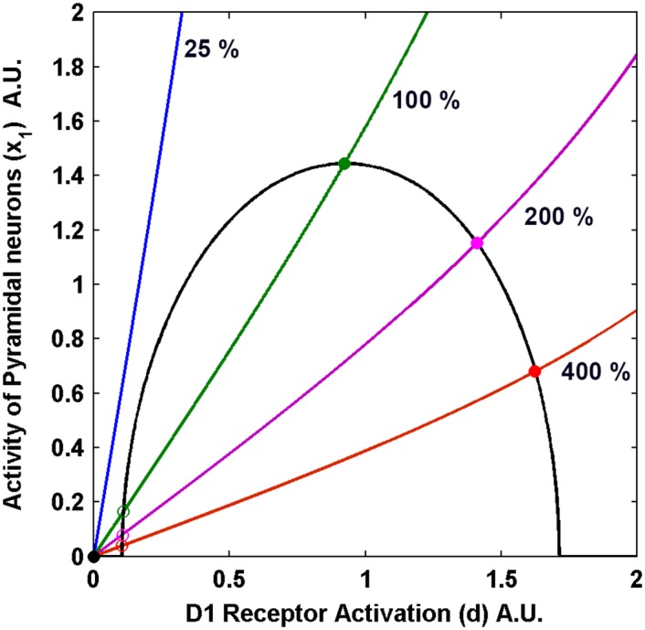
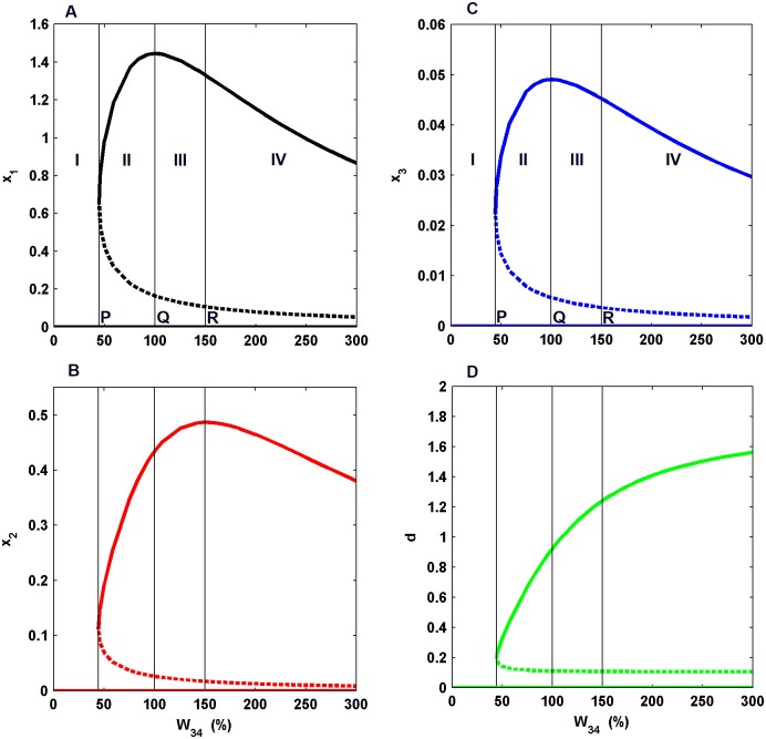
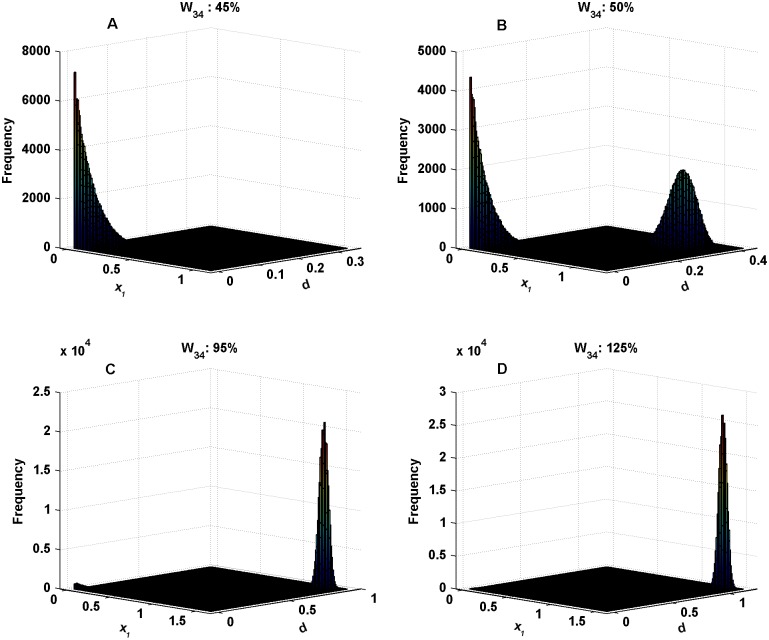
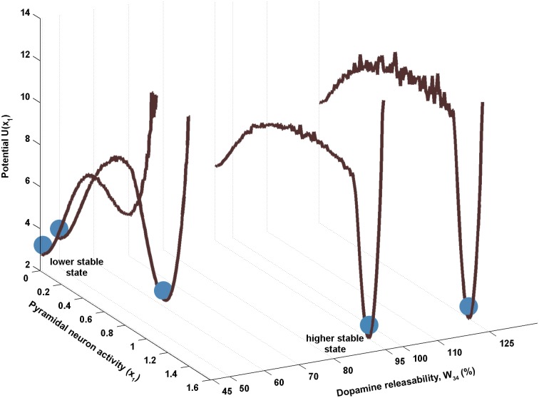
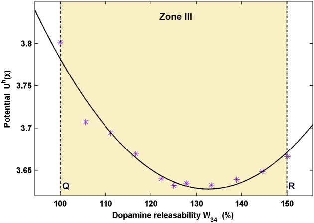
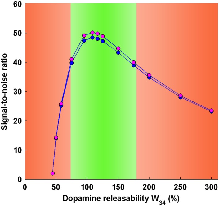
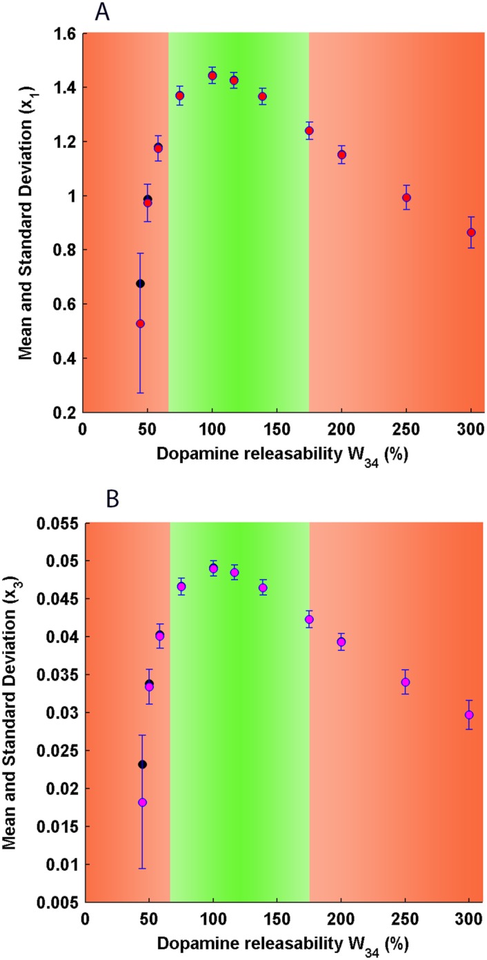
The role of prefronto-mesoprefrontal system in the dopaminergic modulation of working memory during delayed response tasks is well-known. Recently, a dynamical model of the closed-loop mesocortical circuit has been proposed which employs a deterministic framework to elucidate the system's behavior in a qualitative manner. Under natural conditions, noise emanating from various sources affects the circuit's functioning to a great extent. Accordingly in the present study, we reformulate the model into a stochastic framework and investigate its steady state properties in the presence of constant background noise during delay-period. From the steady state distribution, global potential landscape and signal-to-noise ratio are obtained which help in defining robustness of the circuit dynamics. This provides insight into the robustness of working memory during delay-period against its disruption due to background noise. The findings reveal that the global profile of circuit's robustness is predominantly governed by the level of D1 receptor activity and high D1 receptor stimulation favors the working memory-associated sustained-firing state over the spontaneous-activity state of the system. Moreover, the circuit's robustness is further fine-tuned by the levels of excitatory and inhibitory activities in a way such that the robustness of sustained-firing state exhibits an inverted-U shaped profile with respect to D1 receptor stimulation. It is predicted that the most robust working memory is formed possibly at a subtle ratio of the excitatory and inhibitory activities achieved at a critical level of D1 receptor stimulation. The study also paves a way to understand various cognitive deficits observed in old-age, acute stress and schizophrenia and suggests possible mechanistic routes to the working memory impairments based on the circuit's robustness profile.


Conflict of interest statement

Competing Interests: The authors have declared that no competing interests exist.

Figures

Fig 1
Fig 1. Schematic depiction of the Mesocortical circuit.
(A) The interactions of the DLPFC and VTA in the central nervous system. DLPFC (grey region) innervates VTA nuclei (pink) located in the midbrain through corticomesencephalic glutamatergic projections (red). Reciprocally, VTA sends dopaminergic mesocortical projections (blue) to the DLPFC. (B) Detailed connection profile of various functioning modules in the mesocortical circuit. In the DLPFC, the group of pyramidal neurons, x 1 (red), excites itself through recurrent connections, with synaptic strength W 11, as well as excites the group of GABAergic interneurons, x 2 (blue), with synaptic strength W 12. In turn, the GABAergic interneurons, x 2, exert a feed-back inhibition on the activity of pyramidal neurons, x 1, with synaptic strength W 21. Further, excitatory glutamatergic projections from pyramidal neurons, x 1, excite the group of dopaminergic neurons, x 3 (pink), with the efficacy W 13. On excitation, the dopaminergic projections from dopaminergic neurons, x 3, release dopamine (dark red) in the DLPFC with the dopamine releasability W 34. The dopamine pool in the DLPFC leads to D1 receptor stimulation and, accordingly, modulates the various parameters involved in cortical dynamics. The two parameters W 13 (efficacy of cortico-mesencephalic projections) and W 34 (dopamine releasability) govern the efficiency of cross-talk between DLPFC and VTA, and are shown as critical tuning knobs which can be easily rotated to different levels to observe their cumulative effect on the whole circuit dynamics.
Fig 2
Fig 2. Nullcline Plots of the Pyramidal Neurons Activity, x 1 versus D1 Receptor Activation, d for different values of dopamine releasability, W 34.
W 34 is indicated by the % values relative to 0.36. The solid (open) circles represent stable (unstable) fixed points. The intersection points of the curves are the points of global equilibrium of the system, for that particular W 34. The parameters used here are τ1=20,τ2*=6.8,τ3=10,τ4=800 and W11*=0.5588, W12*=0.786, W 13 = 0.023, W 21 = 0.339, a = 0.15.
Fig 3
Fig 3. Bifurcation Diagrams.
These curves depict the equilibrium activity of the pyramidal neurons (A), inhibitory neurons (B), dopaminergic neurons (C) and D1 receptor activation (D) versus the bifurcation parameter W 34. The solid (dotted) lines portray stable (unstable) states of the system. The curves are partitioned into four regions I, II, III and IV. Point P, Q and R are the boundary points of these regions. Point P is the point at which bistability appears in the system. Point Q is the point of maximum equilibrium activity of excitatory neurons and Point R is the point of maximum equilibrium activity of interneurons.
Fig 4
Fig 4. Steady state frequency distributions for different values of dopamine releasability, W 34.
These distributions are obtained by simulating Eqs (7a)–(7d). With increasing W 34, the curves exhibit a gradual transition of relative sample distributions from lower to higher stable state. Additive noise strengths used here are σ 1 = 0.05, σ 2 = 0.01, σ 3 = 0.001, σ 4 = 0.05, estimated in accordance with the scales of magnitude acquired by the different variables in the circuit dynamics.
Fig 5
Fig 5. Global potential landscape w.r.t the pyramidal neuron activity x 1, for different values of DA releasability W 34.
The profiles are obtained from the steady state distributions in variable x 1 for different values of W 34. Here, the lower stable state represents the spontaneous-activity state and the higher stable state represents the sustained-firing state (working memory state) of the system. The system has been depicted by a ball sitting in the potential wells belonging to the different activity states. At low W 34, the system (ball) prefers to rest in the well associated with the lower stable state as it is relatively more robust in comparison to its corresponding higher stable state. However, with the increase in W 34, the higher stable state gradually becomes more robust relative to the corresponding lower stable state.
Fig 6
Fig 6. Profile of variation in potential of higher stable state with change in dopamine releasability W 34.
The points shown in asterisks denote the potentials of higher stable states for the respective dopamine releasability, W 34 in zone III obtained from the global potential landscape. The solid curve is a fitting to the data. The potential is minimum in the region between the points Q and R of maximum equilibrium excitatory and inhibitory activities, respectively. The point of minima represents the maximum robustness that the circuit dynamics may achieve during the delay period. This further suggests the existence of a subtle ratio of the equilibrium excitatory to inhibitory activity which fine tunes the circuit’s robustness and leads to an inverted-U shaped profile of the robustness of higher stable states during the delay period.
Fig 7
Fig 7. Signal-to-noise ratio (SNR) in pyramidal and midbrain activities in the working memory state during delay interval.
The SNR profiles of pyramidal and midbrain activities exhibit an inverted-U shaped profile similar to the signal profile seen in the bifurcation diagram. The SNR profiles are consistent with the error bar plots of pyramidal and midbrain activities, respectively, during delay interval. The optimal region (green zone) is associated with maximum signal-to-noise ratio and signifies establishment of efficient working memory during delay interval. At abnormal levels of dopamine releasability W 34 (right and left, red zone), there occurs a significant decline in the signal-to-noise ratio which may lead to working memory impairment.
Fig 8
Fig 8. Error bar plots of pyramidal (A) and dopaminergic midbrain (B) activity under stochastic mesocortical dynamics during delay interval.
The solid red (magenta) circles represent working memory-associated mean pyramidal (midbrain) activities and the blue error bars denote standard deviations around the mean activities at different dopamine releasability W 34, in the presence of noisy circuit dynamics. The solid black circles represent pyramidal (midbrain) activities obtained from deterministic mesocortical dynamics and constitutes the signal profile. In the optimal region (green zone), the mean pyramidal (midbrain) activity is associated with least noisy fluctuations and coincides with the pyramidal (midbrain) activity of the signal profile. In the region of low dopamine releasability (left, red zone), the pyramidal (midbrain) activity shows dramatic fluctuations such that the mean activity significantly deviates away from the sustained-firing state and shifts towards the spontaneous-activity state. In the region of high dopamine releasability (right, red zone), there does not occur a noticeable deviation of the mean pyramidal (midbrain) activity from the sustained firing state, but, indeed contains significant fluctuations as compared to the optimal region. As evident, the pyramidal and midbrain activity demonstrates a tightly-linked response with regard to their relative noise content.

Similar articles

Cited by

References

    1. Arnsten AF, Wang MJ, Paspalas CD (2012) Neuromodulation of thought: flexibilities and vulnerabilities in prefrontal cortical network synapses. Neuron 76, 223–239. 10.1016/j.neuron.2012.08.038 - DOI - PMC - PubMed
    1. Baddeley A (1992) Working memory. Science 255, 556–559. 10.1126/science.1736359 - DOI - PubMed
    1. Postle BR (2006) Working memory as an emergent property of the mind and brain. Neuroscience 139, 23–38. 10.1016/j.neuroscience.2005.06.005 - DOI - PMC - PubMed
    1. Leung HC, Gore JC, Goldman-Rakic PS (2002) Sustained mnemonic response in the human middle frontal gyrus during on-line storage of spatial memoranda. J Cogn Neurosci 14, 659–671. 10.1162/08989290260045882 - DOI - PubMed
    1. Rowe JB, Toni I, Josephs O, Frackowiak RS, Passingham RE (2000) The prefrontal cortex: response selection or maintenance within working memory? Science 288, 1656–1660. 10.1126/science.288.5471.1656 - DOI - PubMed

Publication types

Substances

LinkOut - more resources