Review
. 2015 Dec 4;8(1):a018903.
doi: 10.1101/cshperspect.a018903.

Maturation and Functional Integration of New Granule Cells into the Adult Hippocampus

Affiliations
Review

Maturation and Functional Integration of New Granule Cells into the Adult Hippocampus

Nicolas Toni et al. Cold Spring Harb Perspect Biol. .

Abstract

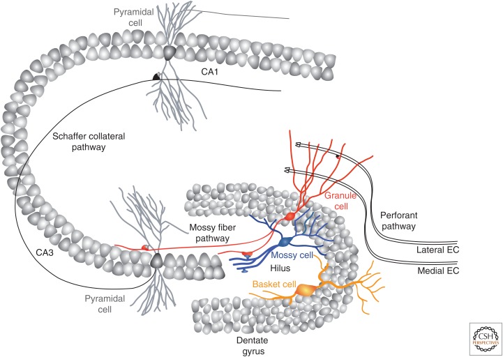
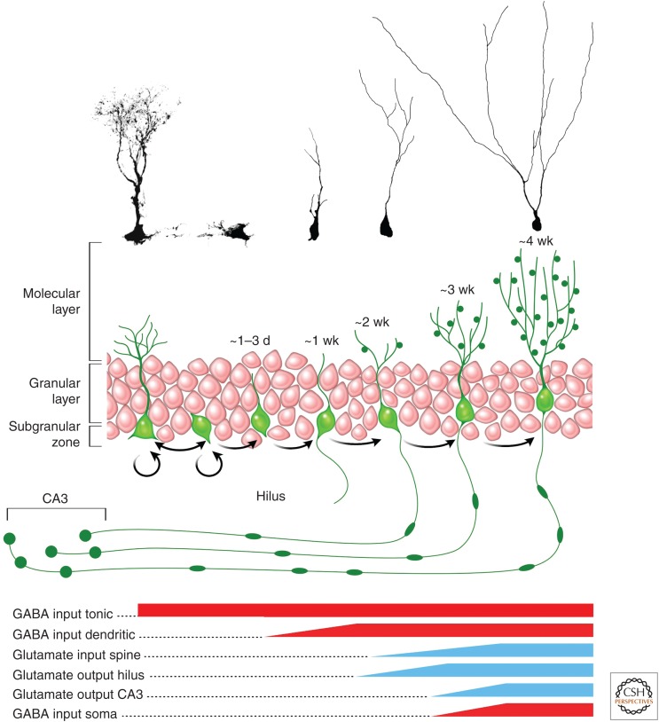
The adult hippocampus generates functional dentate granule cells (GCs) that release glutamate onto target cells in the hilus and cornus ammonis (CA)3 region, and receive glutamatergic and γ-aminobutyric acid (GABA)ergic inputs that tightly control their spiking activity. The slow and sequential development of their excitatory and inhibitory inputs makes them particularly relevant for information processing. Although they are still immature, new neurons are recruited by afferent activity and display increased excitability, enhanced activity-dependent plasticity of their input and output connections, and a high rate of synaptogenesis. Once fully mature, new GCs show all the hallmarks of neurons generated during development. In this review, we focus on how developing neurons remodel the adult dentate gyrus and discuss key aspects that illustrate the potential of neurogenesis as a mechanism for circuit plasticity and function.


Figures

Figure 1.
Figure 1.
Circuits of the hippocampus. Schematic view of a transversal slice through the hippocampus depicting the dentate gyrus, cornus ammonis (CA)3, and CA1. The principal cells located in densely packed layers are synaptically connected via the so-called trisynaptic circuit. The main entry site for this circuit is the perforant path, composed of axons originating from lateral and medial entorhinal cortex (EC) and contacting the outer and medial molecular layer, respectively. Granule neurons then project mossy fibers to CA3 pyramidal neurons, which then project Schaffer collaterals to CA1 pyramidal neurons. Other input to granule neurons include interneurons located in the molecular layer and in the hilus, principally composed of basket and mossy cells.
Figure 2.
Figure 2.
Developmental stages of adult-born granule cells (GCs). Schematic representation and confocal micrographs of the different stages of maturation, from stem cell to mature neuron. The bottom panel indicates the approximate timeline of the major input and output (based on data in Toni and Sultan 2011). CA, Cornus ammonis; GABA, γ-aminobutyric acid.
Figure 3.
Figure 3.
Synaptogenesis in new GCs. (AC) Afferent synapses from perforant path axons. (A) Electron micrographs of dark immunolabeled dendritic spines from new GCs contacting a multiple synapse bouton (left) or a single synapse bouton (right). (B) Three-dimensional reconstructions from serial-section electron micrographs of dendritic protrusions from new GCs (in green). Left panel: filopodia approaching and making a physical contact with a synapse. Middle panel: dendritic spine synapsing with a multiple synapse bouton. Right panel: dendritic spine synapsing with a single synapse bouton. (C) Schematic representation of the hypothesis on the development of dendritic spine connectivity: Left panel: filopodia from new GCs (green) grow preferentially toward preexisting synapses. On stabilization, some filopodia transform into dendritic spines, thereby forming multiple synapse boutons (middle panel). Over maturation time, multiple synapse boutons transform into single-synapse boutons, probably through the retraction of the preexisting spine. Green, dendritic protrusion from new GCs; red, dendritic spine from other GCs; blue, axonal boutons (based on Toni and Sultan 2011). (DF) Efferent synapses on mossy-fiber terminals in the CA3. (D) Electron micrographs of immunolabeled mossy fiber terminals from new GCs at 17 dpi (left panel) and 30 dpi (right panel). Note the thorny excrescence protruding into the terminal on the right panel. (E) Three-dimensional reconstructions of terminals from new GCs synapsing directly on the dendrite (left panel), with a shared thorny excrescence with another, nonimmunolabeled GCs (middle panel) or with an individual thorny excrescence (right panel). Green, terminal from a new GC; red, dendrite from a CA3 pyramidal neuron; blue, terminal from a nonimmunolabeled GC. (F) Schematic depicting the hypothetical development of output connectivity from new GCs. Left panel: the terminal of a new GC synapses first with the dendrite of a CA3 pyramidal cell. Middle panel: on maturation (30 dpi), some thorny excrescences are shared between new and preexisting terminals. Right panel: after 75 dpi, all terminals from new GCs synapse with individual thorny excrescences (based on Toni and Sultan 2011). Scale bars, 1 µm.
Figure 4.
Figure 4.
Functional significance of immature granule cells (GCs). (A–D) Experiments in acute hippocampal slices. (A) A stimulating electrode was placed in the perforant pathway. Stimulation of the medial perforant path (mPP) evoked monosynaptic excitation and disynaptic inhibition via γ-aminobutyric acid (GABA)ergic interneurons (INs) onto GCs. Spiking and the underlying synaptic currents were recorded in individual cells by loose patch followed by whole cell. (B) Example curves of spiking probability versus input strength with example traces. (C) Cumulative distributions of input strength required to elicit 50% spiking probability (activated cells). (D) Activation of GCs evoked by trains and raster plots depicting spikes recorded in response 10 pulses at 10 Hz (five trials/neuron). (From Marin-Burgin et al. 2012; modified, with permission, from The American Association for the Advancement of Science © 2012.) (E,F) In vivo single unit recording in the rat dentate gyrus. Examples of cells with single (E) and multiple (F) place fields. Rate maps and plots showing spikes (Kunze et al. 2009) from the cell superimposed on the rat’s trajectory (gray) are shown in the left and right columns. For the rate maps, blue represents no firing and red represents peak firing (data kindly provided by J.P. Neunuebel and J.J. Knierim).

References

    1. Acsady L, Kamondi A, Sik A, Freund T, Buzsaki G. 1998. GABAergic cells are the major postsynaptic targets of mossy fibers in the rat hippocampus. J Neurosci 18: 3386–3403. - PMC - PubMed
    1. Amaral DG. 1978. A Golgi study of cell types in the hilar region of the hippocampus in the rat. J Comp Neurol 182: 851–914. - PubMed
    1. Amaral DG, Ishizuka N, Claiborne B. 1990. Neurons, numbers and the hippocampal network. Prog Brain Res 83: 1–11. - PubMed
    1. Amaral DG, Scharfman HE, Lavenex P. 2007. The dentate gyrus: Fundamental neuroanatomical organization (dentate gyrus for dummies). Prog Brain Res 163: 3–22. - PMC - PubMed
    1. Barria A, Malinow R. 2005. NMDA receptor subunit composition controls synaptic plasticity by regulating binding to CaMKII. Neuron 48: 289–301. - PubMed

Publication types