. 2016 Mar 4;291(10):5234-46.
doi: 10.1074/jbc.M115.694307. Epub 2015 Dec 4.

Structural Basis of Stereospecificity in the Bacterial Enzymatic Cleavage of β-Aryl Ether Bonds in Lignin

Affiliations

Structural Basis of Stereospecificity in the Bacterial Enzymatic Cleavage of β-Aryl Ether Bonds in Lignin

Kate E Helmich et al. J Biol Chem. .

Abstract

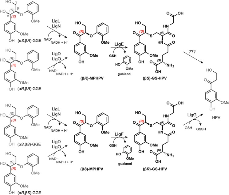
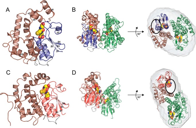
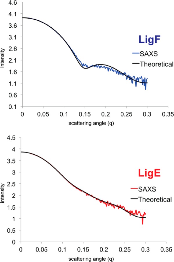
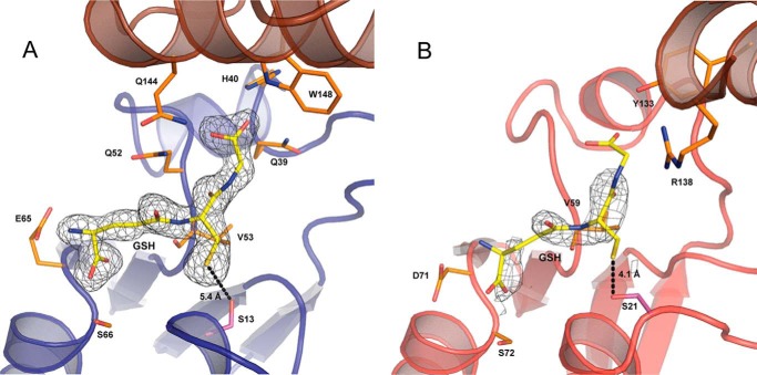
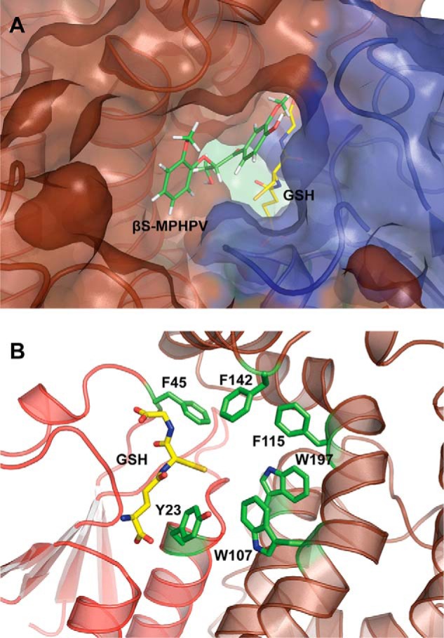
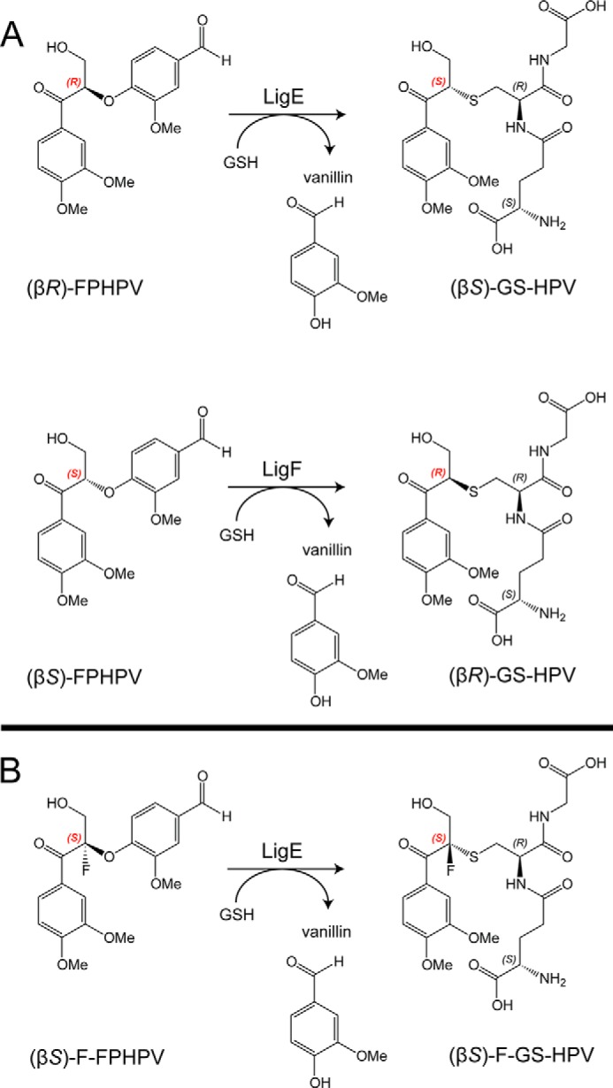
Lignin is a combinatorial polymer comprising monoaromatic units that are linked via covalent bonds. Although lignin is a potential source of valuable aromatic chemicals, its recalcitrance to chemical or biological digestion presents major obstacles to both the production of second-generation biofuels and the generation of valuable coproducts from lignin's monoaromatic units. Degradation of lignin has been relatively well characterized in fungi, but it is less well understood in bacteria. A catabolic pathway for the enzymatic breakdown of aromatic oligomers linked via β-aryl ether bonds typically found in lignin has been reported in the bacterium Sphingobium sp. SYK-6. Here, we present x-ray crystal structures and biochemical characterization of the glutathione-dependent β-etherases, LigE and LigF, from this pathway. The crystal structures show that both enzymes belong to the canonical two-domain fold and glutathione binding site architecture of the glutathione S-transferase family. Mutagenesis of the conserved active site serine in both LigE and LigF shows that, whereas the enzymatic activity is reduced, this amino acid side chain is not absolutely essential for catalysis. The results include descriptions of cofactor binding sites, substrate binding sites, and catalytic mechanisms. Because β-aryl ether bonds account for 50-70% of all interunit linkages in lignin, understanding the mechanism of enzymatic β-aryl ether cleavage has significant potential for informing ongoing studies on the valorization of lignin.

Keywords: X-ray crystallography; enzyme catalysis; enzyme mechanism; enzyme structure; lignin degradation; plant cell wall; protein structure; stereoselectivity; structural enzymology.


Figures

FIGURE 1.
FIGURE 1.
The Sphingobium sp. strain SYK-6 β-etherase pathway. Chiral carbons at which stereospecific reactions occur are highlighted (red). Stereospecific reactions for (αSR)-GGE and (αSS)-GGE oxidation (by LigL and LigN) and for (αRR)-GGE and (αRS)-GGE oxidation (by LigD and LigO), the GSH-dependent stereospecific cleavage reactions of (βR)-MPHPV (by LigE) and (βS)-MPHPV (by LigF), and the stereospecific lyase reaction of LigG with (βS)-GS-HPV are shown.
FIGURE 2.
FIGURE 2.
LigE and LigF structures. A, schematic representation of LigF, including the N-terminal thioredoxin domain (blue), the C-terminal α-helical domain (brown), and the short linker (gray). Bound GSH is shown as yellow spheres. B, schematic representation of the LigF dimer with the proposed substrate binding site (Fig. 5A) circled. C, schematic representation of LigE, including the N-terminal thioredoxin domain (red), the C-terminal α-helical domain (brown), and the short linker (gray). Bound GSH is shown as yellow spheres. D, schematic representation of the dimer of LigE with the proposed binding site (Fig. 5B) circled.
FIGURE 3.
FIGURE 3.
Theoretical and experimental small angle x-ray scattering scatter curves. The scattering angle (q) versus the intensity of the scattering plots shows the experimentally observed data and the theoretical scattering determined using CRYSOL from the x-ray structures of the dimers. LigF is shown at the top in blue, and LigE is shown at the bottom in red.
FIGURE 4.
FIGURE 4.
Representative cytosolic GST dimer forms. Representatives from several GST classes are shown, in which one molecule of the dimer is shown in brown, the second molecule of the dimer is shown in rainbow colors (N terminus in blue to C terminus in red), and the bound glutathione or glutathione analog is shown as yellow spheres. The Alpha (Protein Data Bank entry 1GUH; human GST A1-1), Mu (2GST; rat), Pi (2GSR; pGST P1-1 from pig), Sigma (1GSQ; squid), Theta (1LJR; human hGST T2-2), Beta (2PMT; bacterial GST from P. mirabilis), Omega (3LFL; human GST Omega-1), and LigG (4G10; Sphingobium sp. SYK-6) dimers show variations on the α3/α4 canonical four-helix bundle dimer structure, whereas the GSTFuA structure from P. chrysosporium shows a non-canonical dimer formed via interaction between α4 and the C-terminal domain of the second molecule of the dimer.
FIGURE 5.
FIGURE 5.
Glutathione binding sites in LigF and LigE. A, the GSH binding site in LigF is located in a cleft between the thioredoxin and α-helical domains. Density for the bound GSH (yellow sticks) is shown in gray contoured to 1.0σ (CC = 0.97). Residues interacting with the γ-glutamyl (Glu-65 and Ser-66), cysteinyl (Gln-52 and Val-53), and glycine (Gln-144, His-40, Trp-148, and Gln-39) residues of the bound GSH are shown as orange sticks. The distance between the GSH sulfur and the active site serine 13 (purple sticks) is 5.4 Å. B, the GSH binding in LigE is located on a surface-exposed face between the thioredoxin and α-helical domains. Density for the bound GSH (yellow sticks) from chain A of the model is shown in gray contoured to 1.0σ (CC = 0.71). Residues interacting with the γ-glutamyl (Asp-71 and Ser-72), cysteinyl (Val-59), and glycine (Arg-138 and Tyr-133) of the GSH are shown as orange sticks. The distance between the GSH sulfur and the active site serine 21 (purple sticks) is 4.1 Å.
FIGURE 6.
FIGURE 6.
Substrate binding sites in LigF and LigE. A, model of ternary complex LigFΔ242-GSH·(βS)-MPHPV. Schematic representations are shown of the N-terminal thioredoxin domain (blue) and the C-terminal α-helical domain (brown) with the circled region from Fig. 2B detailed in a transparent surface rendering. The bound glutathione (yellow) and docked (βS)-MPHPV (green) are shown as sticks. B, proposed substrate binding surface in LigE. Schematic representations are shown for the LigE dimer, and the circled region from Fig. 2D is detailed, showing the hydrophobic aromatic substrate binding pocket formed by Phe-45, Phe-142, Phe-115, Trp-197, Trp-107, and Tyr-23 as green sticks.
FIGURE 7.
FIGURE 7.
A, structure of an MPHPV analog substrate, FPHPV, that was used in the LigE- and LigF-catalyzed reactions, converting FPHPV to vanillin and GS-HVP. B, LigE-catalyzed β-ether elimination reaction with fluorinated model substrate (βS)-F-FPHPV, resulting in formation of vanillin and (βS)-F-GS-HVP.
FIGURE 8.
FIGURE 8.
LigE and LigF pH rate profile. The effect of pH on β-etherase activities was determined for each enzyme, revealing that LigE (triangles), LigF (circles), LigFΔ242 (diamonds), and LigFΔ242-S13A (squares) have pH optima at pH 8.0. Plotted as a function of pH (x axis) are the specific enzymatic activities (y axis) of β-etherases with either (βR)-FPHPV (LigE) or (βS)-FPHPV (LigF, LigFΔ242, and LigFΔ242-S13A) as the assay substrate (1.5 mm initial concentration). Error bars, S.D. of triplicate measurements.

References

    1. Simmons B. A., Loque D., and Blanch H. W. (2008) Next-generation biomass feedstocks for biofuel production. Genome Biol. 9, 242. - PMC - PubMed
    1. Bugg T. D. H., Ahmad M., Hardiman E. M., and Rahmanpour R. (2011) Pathways for degradation of lignin in bacteria and fungi. Nat. Prod. Rep. 28, 1883–1896 - PubMed
    1. Bugg T. D. H., Ahmad M., Hardiman E. M., and Singh R. (2011) The emerging role for bacteria in lignin degradation and bio-product formation. Curr. Opin. Biotechnol. 22, 394–400 - PubMed
    1. Leonowicz A., Cho N. S., Luterek J., Wilkolazka A., Wojtas-Wasilewska M., Matuszewska A., Hofrichter M., Wesenberg D., and Rogalski J. (2001) Fungal laccase: properties and activity on lignin. J. Basic Microbiol. 41, 185–227 - PubMed
    1. Martínez A. T., Speranza M., Ruiz-Dueñas F. J., Ferreira P., Camarero S., Guillén F., Martínez M. J., Gutiérrez A., and del Río J. C. (2005) Biodegradation of lignocellulosics: microbial chemical, and enzymatic aspects of the fungal attack of lignin. Int. Microbiol. 8, 195–204 - PubMed

Publication types