

doi: 10.1128/JVI.02779-15.
Print 2016 Feb 15.
A Highly Conserved Residue of the HIV-1 gp120 Inner Domain Is Important for Antibody-Dependent Cellular Cytotoxicity Responses Mediated by Anti-cluster A Antibodies
Affiliations
- PMID: 26637462
- PMCID: PMC4733974
- DOI: 10.1128/JVI.02779-15
Item in Clipboard
A Highly Conserved Residue of the HIV-1 gp120 Inner Domain Is Important for Antibody-Dependent Cellular Cytotoxicity Responses Mediated by Anti-cluster A Antibodies
J Virol.
.
Abstract
Previous studies have shown that sera from HIV-1-infected individuals contain antibodies able to mediate antibody-dependent cellular cytotoxicity (ADCC). These antibodies preferentially recognize envelope glycoprotein (Env) epitopes induced upon CD4 binding. Here, we show that a highly conserved tryptophan at position 69 of the gp120 inner domain is important for ADCC mediated by anti-cluster A antibodies and sera from HIV-1-infected individuals.
Copyright © 2016, American Society for Microbiology. All Rights Reserved.
Figures
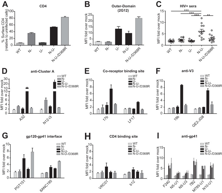
Effect of Nef, Vpu, and Env-CD4 interaction on recognition of infected cells by HIV+ sera and a panel of monoclonal antibodies. CEM.NKr cells infected with a panel of vesicular stomatitis virus glycoprotein-pseudotyped NL4.3–GFP ADA viruses expressing wild type (WT) or a CD4-binding site (D368R) Env variant, lacking Nef (N−), Vpu (U−), or both Nef and Vpu (N− U−), were stained at 48 h postinfection with an anti-CD4 antibody (OKT4) (A); 1 μg/ml of the CD4-independent outer domain recognizing 2G12 antibody (B); a 1/1,000 dilution of sera from 12 HIV-1-infected individuals (HIV+ sera) (C); or 1 μg/ml of anti-cluster A (A32 and N12-i3) (D), anti-CoRBS (17b and LF17) (E), anti-V3 (19b and GE2-JG8) (F), anti-gp120-gp41 interface (PGT151 and 8ANC195) (G), anti-CD4-binding site (VRC01 and b12) (H), and anti-gp41 (F240, N5-U1, N5-U3, 7B2, M785-U1, and N10-U1) (I) antibodies and then fluorescently labeled with an Alexa Fluor 647-conjugated anti-human IgG secondary Ab. Shown is the fold increase of staining relative to mock for all tested sera and antibodies. Data shown are the results of 3 different experiments, and error bars depict the standard errors of the means. Statistical significance was tested using paired one-way analyses of variance (**, P < 0.01; ***, P < 0.001). MFI, mean fluorescence intensity.
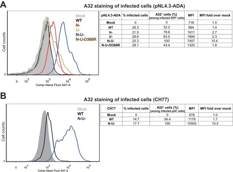
Recognition of HIV-1-infected cells by the anti-cluster A32 antibody. CEM.NKr cells infected with a panel of vesicular stomatitis virus glycoprotein-pseudotyped NL4.3–GFP ADA viruses expressing wild type (WT) or a CD4-binding site (D368R) Env variant, lacking Nef (N−), Vpu (U−), or both Nef and Vpu (N− U−) (A), or with vesicular stomatitis virus glycoprotein-pseudotyped primary T/F CH77 infectious molecular clone (B) were stained at 48 h postinfection with the anti-cluster A A32 antibody (1 μg/ml) and then fluorescently labeled with an Alexa Fluor 647-conjugated anti-human IgG secondary Ab. Histograms depicting representative staining of infected (GFP+ [A] or p24+ [B]) cells are shown. Right panels show the percentages of infected cells, the percentages of infected (GFP+ [A] or p24+ [B]) cells that were recognized by A32, and the mean fluorescence intensity (MFI) of these cells. Mean fluorescence intensity of infected cells over that of mock-infected cells is shown in the last column.
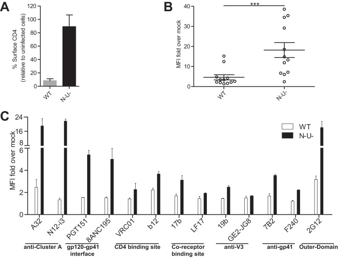
Effect of Nef and Vpu on recognition of cells infected with a primary isolate by HIV+ sera and a panel of monoclonal antibodies. (A and B) CEM.NKr cells infected with vesicular stomatitis virus glycoprotein-pseudotyped T/F CH77 expressing wild type (WT) or lacking Nef and Vpu (N− U−) were stained at 48 h postinfection with an anti-CD4 antibody (OKT4) (A) or a 1/1,000 dilution of sera from 12 HIV-1-infected individuals (HIV+ sera) (B). (C) CH77-infected cells were also stained with a panel of anti-gp120 and -gp41 antibodies (1 μg/ml) and then fluorescently labeled with an Alexa Fluor 647-conjugated anti-human IgG secondary Ab. Data shown are the results of 3 independent experiments, with medians ± interquartile ranges. Statistical significance was tested using paired one-way analyses of variance (***, P < 0.001).
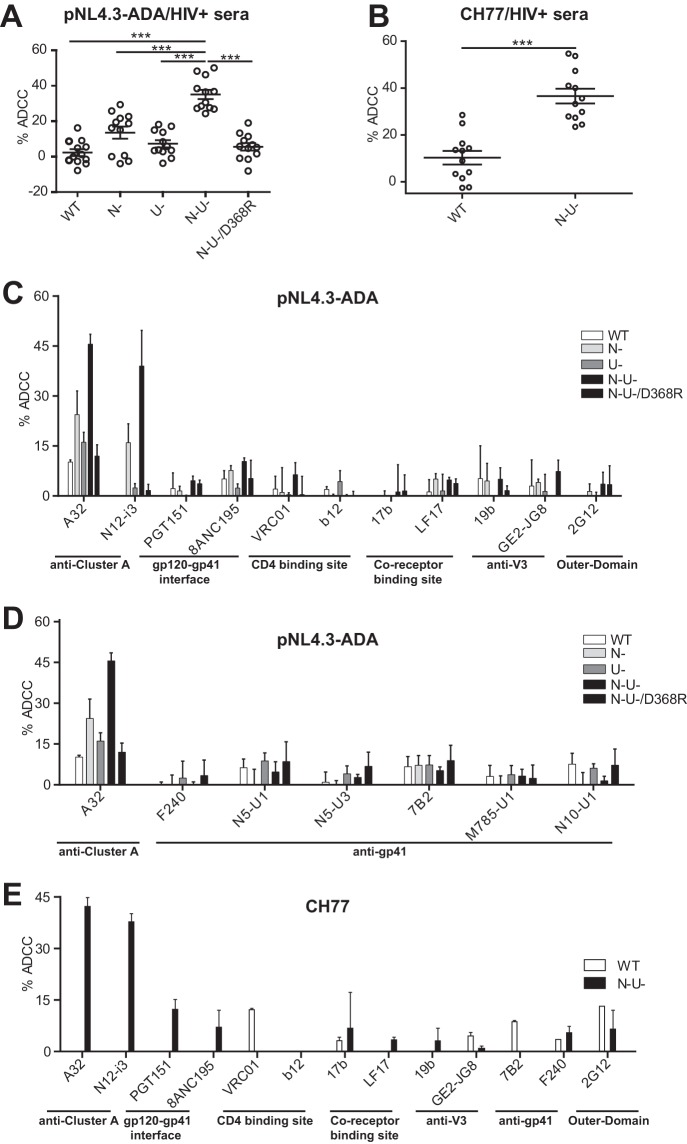
Effect of Nef, Vpu, and Env-CD4 interaction on ADCC responses mediated by HIV+ sera and a panel of monoclonal antibodies. (A) CEM.NKr cells infected with a panel of vesicular stomatitis virus glycoprotein-pseudotyped NL4.3–GFP ADA viruses expressing wild type (WT) or a CD4-binding site (D368R) Env variant, lacking Nef (N−), Vpu (U−), or both Nef and Vpu (N− U−), were used at 48 h postinfection as target cells in our FACS-based ADCC assay (5) to determine their susceptibility to ADCC mediated by a 1/1,000 dilution of sera from 12 HIV-1-infected individuals. (B) The susceptibility of vesicular stomatitis virus glycoprotein-pseudotyped T/F CH77-infected cells expressing wild type (WT) or lacking Nef and Vpu (N− U−) to ADCC mediated by the same panel of HIV+ sera was also evaluated. (C and D) The susceptibility of pNL4.3-ADA-infected cells to ADCC mediated by 5 μg/ml of anti-gp120 (anti-cluster A antibodies A32 and N12-i3; anti-CD4-binding site antibodies VRC01 and b12; anti-CoRBS antibodies 17b and LF17; anti-V3 antibodies 19b and GE2-JG8; anti-outer domain antibody 2G12) or anti-gp120-gp41 (PGT151 and 8ANC195) (C) or anti-gp41 (F240, N5-U1, N5-U3, 7B2, M785-U1, and N10-U1) (D) antibodies was also evaluated. (E) Susceptibility of CH77-infected cells to anti-gp120 and anti-gp41 antibodies. Peripheral blood mononuclear cells from healthy donors were used as effector cells. Data shown are the results of 3 independent experiments, with medians ± interquartile ranges. Statistical significance was tested using paired one-way analyses of variance (***, P < 0.001).
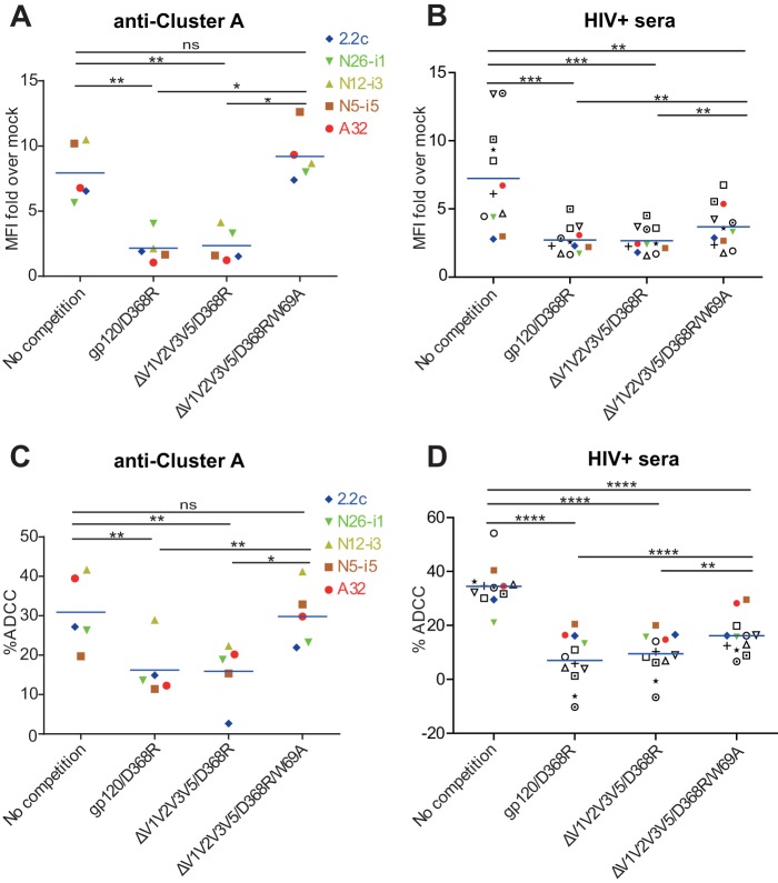
Tryptophan 69 in layer 1 is important for ADCC responses mediated by anti-cluster A antibodies and some HIV+ sera. CEM.NKr cells infected with N− U− vesicular stomatitis virus glycoprotein-pseudotyped NL4-3–GFP ADA virus were used at 48 h postinfection for surface staining (A and B) or FACS-based ADCC assay (C and D) using 5 μg/ml of anti-cluster A antibodies (A32, N5-i5, N12-i3, N26-i1, and 2.2c) or a 1/1,000 dilution of sera from 12 HIV-1-infected individuals (HIV+ sera, different color or symbol for each serum sample) in the absence or presence of recombinant gp120/D368R (10 μg/μg of antibody), ΔV1V2V3V5/D368R (6 μg/μg of antibody), or ΔV1V2V3V5/D368R/W69A (6 μg/μg of antibody) for 30 min at room temperature. Data shown are representative of at least 3 independent experiments. Statistical significance was tested using paired one-way analyses of variance (*, P < 0.05; **, P < 0.01; ***, P < 0.001; ****, P < 0.0001; ns, not significant).
References
-
- Veillette M, Coutu M, Richard J, Batraville LA, Dagher O, Bernard N, Tremblay C, Kaufmann DE, Roger M, Finzi A. 2015. The HIV-1 gp120 CD4-bound conformation is preferentially targeted by antibody-dependent cellular cytotoxicity-mediating antibodies in sera from HIV-1-infected individuals. J Virol 89:545–551. doi:10.1128/JVI.02868-14. - DOI - PMC - PubMed
-
- Batraville LA, Richard J, Veillette M, Labbe AC, Alary M, Guedou F, Kaufmann DE, Poudrier J, Finzi A, Roger M. 2014. Anti-HIV-1 envelope immunoglobulin Gs in blood and cervicovaginal samples of Beninese commercial sex workers. AIDS Res Hum Retroviruses 30:1145–1149. doi:10.1089/aid.2014.0163. - DOI - PMC - PubMed
-
- Richard J, Veillette M, Brassard N, Iyer SS, Roger M, Martin L, Pazgier M, Schon A, Freire E, Routy JP, Smith AB III, Park J, Jones DM, Courter JR, Melillo BN, Kaufmann DE, Hahn BH, Permar SR, Haynes BF, Madani N, Sodroski JG, Finzi A. 2015. CD4 mimetics sensitize HIV-1-infected cells to ADCC. Proc Natl Acad Sci U S A 112:E2687–E2694. doi:10.1073/pnas.1506755112. - DOI - PMC - PubMed
-
- Veillette M, Desormeaux A, Medjahed H, Gharsallah NE, Coutu M, Baalwa J, Guan Y, Lewis G, Ferrari G, Hahn BH, Haynes BF, Robinson JE, Kaufmann DE, Bonsignori M, Sodroski J, Finzi A. 2014. Interaction with cellular CD4 exposes HIV-1 envelope epitopes targeted by antibody-dependent cell-mediated cytotoxicity. J Virol 88:2633–2644. doi:10.1128/JVI.03230-13. - DOI - PMC - PubMed
Publication types
MeSH terms
Substances
Grants and funding
LinkOut - more resources
Full Text Sources
Other Literature Sources
Research Materials
