

Targeted DNA Sequencing from Autism Spectrum Disorder Brains Implicates Multiple Genetic Mechanisms
- PMID: 26637798
- PMCID: PMC4672379
- DOI: 10.1016/j.neuron.2015.11.009
Targeted DNA Sequencing from Autism Spectrum Disorder Brains Implicates Multiple Genetic Mechanisms
Abstract
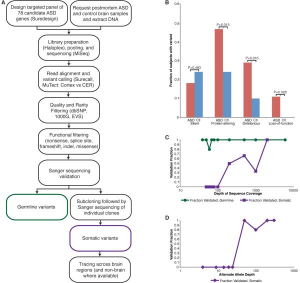
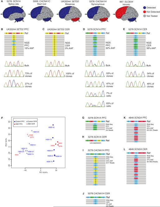
Single nucleotide variants (SNVs), particularly loss-of-function mutations, are significant contributors to autism spectrum disorder (ASD) risk. Here we report the first systematic deep sequencing study of 55 postmortem ASD brains for SNVs in 78 known ASD candidate genes. Remarkably, even without parental samples, we find more ASD brains with mutations that are protein-altering (26/55 cases versus 12/50 controls, p = 0.015), deleterious (16/55 versus 5/50, p = 0.016), or loss-of-function (6/55 versus 0/50, p = 0.028) compared to controls, with recurrent deleterious mutations in ARID1B, SCN1A, SCN2A, and SETD2, suggesting these mutations contribute to ASD risk. In several cases, the identified mutations and medical records suggest syndromic ASD diagnoses. Two ASD and one Fragile X premutation case showed deleterious somatic mutations, providing evidence that somatic mutations occur in ASD cases, and supporting a model in which a combination of germline and/or somatic mutations may contribute to ASD risk on a case-by-case basis.
Copyright © 2015 Elsevier Inc. All rights reserved.
Figures
References
-
- Betancur C. Etiological heterogeneity in autism spectrum disorders: more than 100 genetic and genomic disorders and still counting. Brain research. 2011;1380:42–77. - PubMed
-
- Chen JA, Penagarikano O, Belgard TG, Swarup V, Geschwind DH. The emerging picture of autism spectrum disorder: genetics and pathology. Annual review of pathology. 2015;10:111–144. - PubMed
Publication types
MeSH terms
Substances
Grants and funding
- U01MH106883/MH/NIMH NIH HHS/United States
- R01 MH083565/MH/NIMH NIH HHS/United States
- U01 MH106883/MH/NIMH NIH HHS/United States
- T32 GM007226/GM/NIGMS NIH HHS/United States
- P50MH106934/MH/NIMH NIH HHS/United States
- T32 GM007753/GM/NIGMS NIH HHS/United States
- U01MH106874/MH/NIMH NIH HHS/United States
- P30 HD018655/HD/NICHD NIH HHS/United States
- R01MH083565/MH/NIMH NIH HHS/United States
- HHMI/Howard Hughes Medical Institute/United States
- 5T32 GM007226-39/GM/NIGMS NIH HHS/United States
- T32GM007753/GM/NIGMS NIH HHS/United States
- U01 MH106874/MH/NIMH NIH HHS/United States
- MR/L022656/1/MRC_/Medical Research Council/United Kingdom
- P50 MH106934/MH/NIMH NIH HHS/United States
- R01 NS035129/NS/NINDS NIH HHS/United States
- U01 MH103339/MH/NIMH NIH HHS/United States
- UO1MH103339/MH/NIMH NIH HHS/United States
LinkOut - more resources
Full Text Sources
Other Literature Sources
Medical
Molecular Biology Databases
