. 2015 Dec 3;163(6):1444-56.
doi: 10.1016/j.cell.2015.10.072.

Epithelial IL-18 Equilibrium Controls Barrier Function in Colitis

Affiliations

Epithelial IL-18 Equilibrium Controls Barrier Function in Colitis

Roni Nowarski et al. Cell. .

Abstract

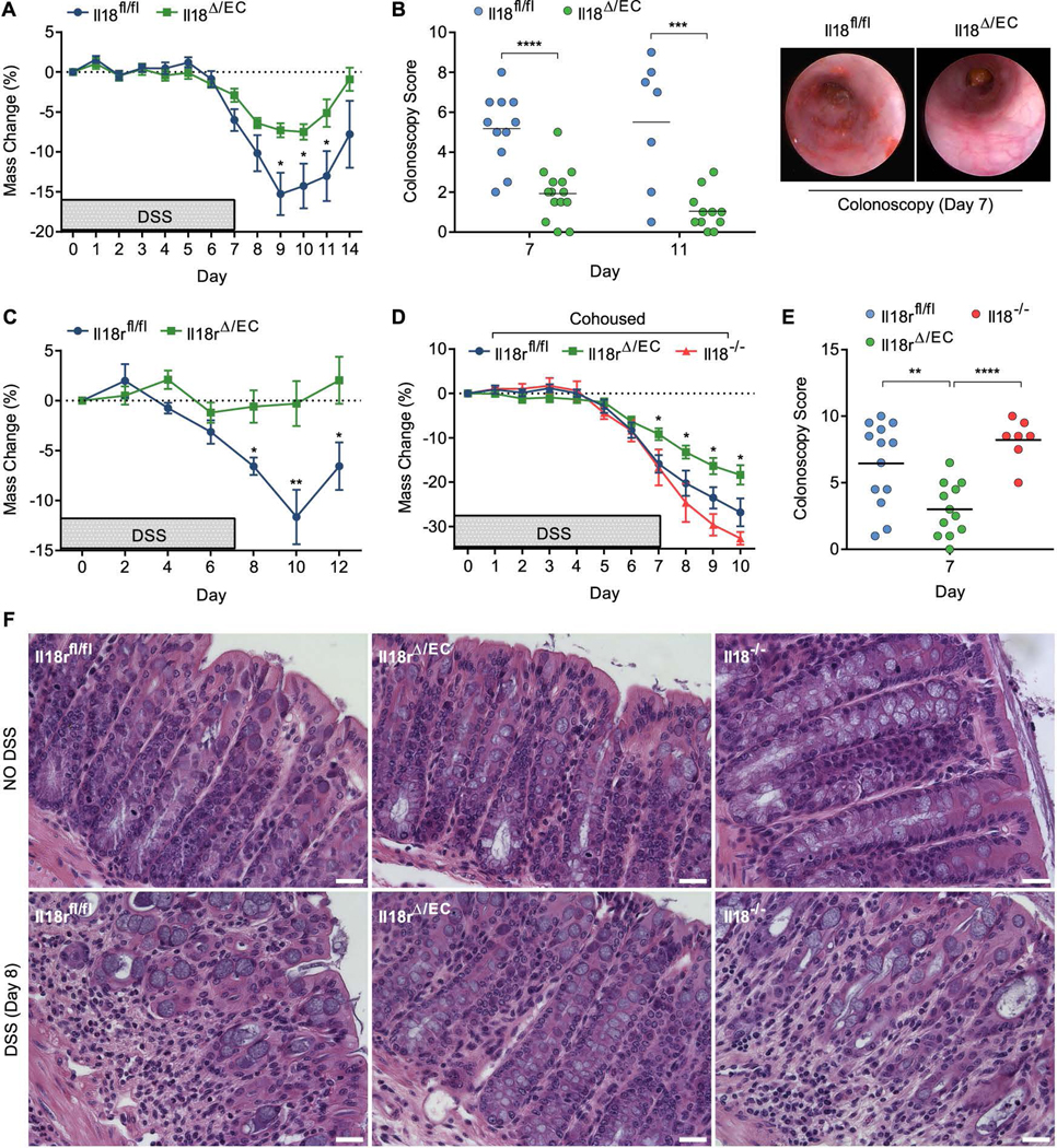
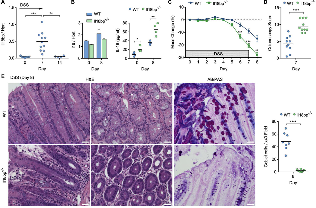
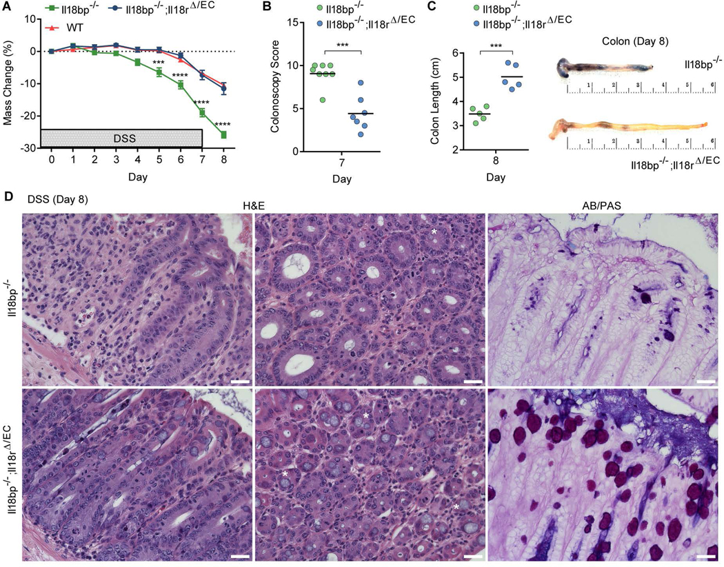
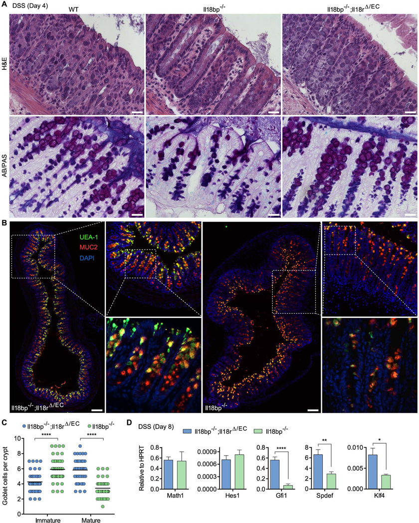
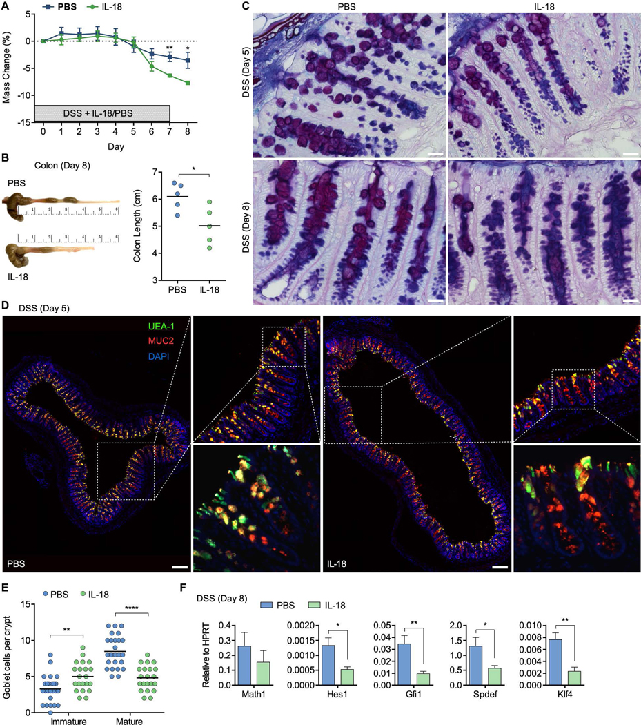
The intestinal mucosal barrier controlling the resident microbiome is dependent on a protective mucus layer generated by goblet cells, impairment of which is a hallmark of the inflammatory bowel disease, ulcerative colitis. Here, we show that IL-18 is critical in driving the pathologic breakdown of barrier integrity in a model of colitis. Deletion of Il18 or its receptor Il18r1 in intestinal epithelial cells (Δ/EC) conferred protection from colitis and mucosal damage in mice. In contrast, deletion of the IL-18 negative regulator Il18bp resulted in severe colitis associated with loss of mature goblet cells. Colitis and goblet cell loss were rescued in Il18bp(-/-);Il18r(Δ/EC) mice, demonstrating that colitis severity is controlled at the level of IL-18 signaling in intestinal epithelial cells. IL-18 inhibited goblet cell maturation by regulating the transcriptional program instructing goblet cell development. These results inform on the mechanism of goblet cell dysfunction that underlies the pathology of ulcerative colitis.


Figures

Figure 1
Figure 1. Epithelial IL-18/IL-18R signaling promotes DSS-induced colitis
(A–F) To induce colitis, mice were administered 2% DSS in drinking water for 7 days. (A) Weight loss of cohoused Il18fl/fl and Il18Δ/EC littermates (n=11–14). (B) Colonoscopy severity score of Il18fl/fl and Il18Δ/EC mice on day 7 and 11 post DSS (left) and representative endoscopic view of the mouse colon on day 7 post DSS (right). (C) Weight loss of cohoused Il18rfl/fl and Il18rΔ/EC littermates (n=8). (D) Littermate Il18rfl/fl and Il18rΔ/EC mice were cohoused with Il18−/− mice for 8 weeks, after which DSS was administered and weight loss recorded (n=7–13). (E) Colonoscopy severity score of cohoused Il18rfl/fl, Il18rΔ/EC and Il18−/− on day 7. (F) Representative H&E staining of distal colon sections obtained from cohoused Il18rfl/fl, Il18rΔ/EC and Il18−/− mice given water (top) or DSS (bottom) and assessed on day 8. Note the disruption of crypt structure and mucosal immune cell infiltration in Il18rfl/fl and Il18−/− mice. Scale bar = 25 μm. Data are represented as mean ± SEM. *, p<0.05; **, p<0.01; ***, p<0.001; ****, p<0.0001 by unpaired Student’s t-test. Related to Figure S1–3.
Figure 2
Figure 2. Hematopoietic/endothelial IL-18, but not IL-18R, promotes DSS-induced colitis
(A) Weight loss of cohoused Il18fl/fl and Il18Δ/HE littermates treated with 2% DSS for 7 days (n=4). (B) Colonoscopy severity score of Il18fl/fl and Il18Δ/HE mice on day 7 and 11. (C) Weight loss of cohoused Il18rfl/fl and Il18rΔ/HE littermates (n=6). (D) Colonoscopy severity score of Il18rfl/fl and Il18rΔ/HE mice on day 7. (E) Representative H&E staining of distal colon sections obtained from cohoused Il18rfl/fl and Il18rΔ/HE mice given water (top) or DSS (bottom) and assessed on day 8. Scale bar = 25 μm. Data are represented as mean ± SEM. *, p<0.05; **, p<0.01; ***, p<0.001; by unpaired Student’s t-test. Related to Figure S1–3.
Figure 3
Figure 3. Hyperactive IL-18 signaling drives colitis and goblet cell loss in Il18bp−/− mice
(A) Wild-type (WT) mice were treated with 2% DSS in drinking water and Il18bp mRNA expression in the distal colon was measured over 14 days. (B) IL-18 mRNA expression in distal colon (left) and protein secretion in colonic explants (right) of il18bp−/− and WT littermates. (C) Weight loss following DSS treatment in cohoused WT and Il18bp−/− littermates (n=10). (D) Colonoscopy severity score of WT and Il18bp−/− mice. (E) Representative H&E and AB/PAS staining of distal colon sections obtained from cohoused WT (top) and Il18bp−/− (bottom) littermates on day 8 post DSS treatment. Asterisks mark example goblet cells. Right, enumeration of goblet cells in histological sections from cohoused WT and Il18bp−/− littermates. Scale bar = 25 μm. Data are represented as mean ± SEM. *, p<0.05; **, p<0.01; ***, p<0.001; ****, p<0.0001 by unpaired Student’s t-test. Related to Figure S1–4.
Figure 4
Figure 4. Deletion of epithelial IL-18R rescues goblet cell loss and colitis in Il18bp−/− mice
(A) Weight loss of cohoused Il18bp−/−, Il18bp−/−;Il18rΔ/EC and WT littermates treated with 2% DSS for 7 days (n=10–14). (B) Colonoscopy severity score of Il18bp−/− and Il18bp−/−;Il18rΔ/EC littermates on day 7. (C) Gross pathology of colons on day 8 post DSS. Note the extensive shortening, bleeding and diarrhea in Il18bp−/− mice. (D) Representative H&E and AB/PAS staining of distal colon sections obtained from cohoused Il18bp−/− (top) and Il18bp−/−;Il18rΔ/EC mice (bottom) on day 8 post DSS treatment. Asterisks mark example goblet cells. Scale bar = 25 μm. Data are represented as mean ± SEM. **, p<0.01; ***, p<0.001; ****, p<0.0001 by unpaired Student’s t-test.
Figure 5
Figure 5. Hyperactive IL-18 signaling prevents goblet cell maturation prior to colitis
(A) Representative H&E and AB/PAS staining of distal colon sections obtained from WT, Il18bp−/− and Il18bp−/−;Il18rΔ/EC littermates on day 4 post DSS treatment prior to onset of colitis. Note reduction in PAS+ mature goblet cells in Il18bp−/− mice. Scale bar = 25 μm. (B) Epifluorescence images (left, top right panels) and confocal stacks (bottom right panels) of distal colon sections obtained on day 4 post DSS and stained with the fucose-binding lectin UEA-1 and anti-MUC2. Scale bar = 150 μm. (C) Enumeration of immature and mature goblet cells in distal colon sections stained as in (B). Immature goblet cells were scored as UEA-1lo/− cells containing low area (<10 μm in diameter) MUC2 staining, and mature goblet cells were scored as cells containing large area (>10 μm in diameter) MUC2+UEA-1bright staining. (D) Distal colon samples were obtained on day 8 post DSS and used for gene expression analysis by qPCR. Data are represented as mean ± SEM. *, p<0.05; ****, p<0.0001 by unpaired Student’s t-test. Related to Figure S4.
Figure 6
Figure 6. IL-18 directly controls goblet cell maturation and colitis
(A) WT mice received daily i.p. injections of 1 μg recombinant IL-18 or PBS during the course of 7 day 2% DSS administration and weight loss was recorded (n=5). (B) Gross pathology of colons on day 8 post DSS. Note reduction in colon length following IL-18 treatment. (C) Representative AB/PAS staining of distal colon sections obtained on day 5 or 8 post DSS treatment from WT littermates receiving recombinant IL-18 or PBS. Note reduction in PAS+ mature goblet cells in mice receiving recombinant IL-18. Scale bar = 25 μm. (D) Epifluorescence images (left, top right panels) and confocal stacks (bottom right panels) of distal colon sections obtained on day 5 post DSS and stained with UEA-1 and anti-MUC2. Scale bar = 150 μm. (E) Enumeration of immature and mature goblet cells in distal colon sections stained as in (D). Immature and mature goblet cells were scored as in Figure 5C. (F) Distal colon samples were obtained on day 8 post DSS and used for gene expression analysis by qPCR. Data are represented as mean ± SEM. *, p<0.05; **, p<0.01; ****, p<0.0001 by unpaired Student’s t-test. Related to Figure S4.

Comment in

References

    1. Adachi O, Kawai T, Takeda K, Matsumoto M, Tsutsui H, Sakagami M, Nakanishi K, Akira S. Targeted disruption of the MyD88 gene results in loss of IL-1- and IL-18-mediated function. Immunity. 1998;9:143–150. - PubMed
    1. Adolph TE, Tomczak MF, Niederreiter L, Ko HJ, Bock J, Martinez-Naves E, Glickman JN, Tschurtschenthaler M, Hartwig J, Hosomi S, et al. Paneth cells as a site of origin for intestinal inflammation. Nature. 2013;503:272–276. - PMC - PubMed
    1. Asquith M, Powrie F. An innately dangerous balancing act: intestinal homeostasis, inflammation, and colitis-associated cancer. J Exp Med. 2010;207:1573–1577. - PMC - PubMed
    1. Bamias G, Corridoni D, Pizarro TT, Cominelli F. New Insights into the Dichotomous Role of Innate Cytokines in Gut Homeostasis and Inflammation. Cytokine. 2012;59:451–459. - PMC - PubMed
    1. Bauernfeind FG, Horvath G, Stutz A, Alnemri ES, MacDonald K, Speert D, Fernandes-Alnemri T, Wu J, Monks BG, Fitzgerald KA, et al. Cutting edge: NF-kappaB activating pattern recognition and cytokine receptors license NLRP3 inflammasome activation by regulating NLRP3 expression. J Immunol. 2009;183:787–791. - PMC - PubMed

Publication types

MeSH terms

Substances