. 2015 Dec 7:5:18132.
doi: 10.1038/srep18132.

Iron-overload injury and cardiomyopathy in acquired and genetic models is attenuated by resveratrol therapy

Affiliations

Iron-overload injury and cardiomyopathy in acquired and genetic models is attenuated by resveratrol therapy

Subhash K Das et al. Sci Rep. .

Abstract

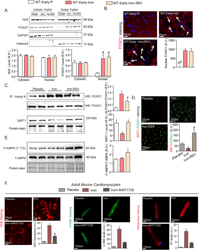
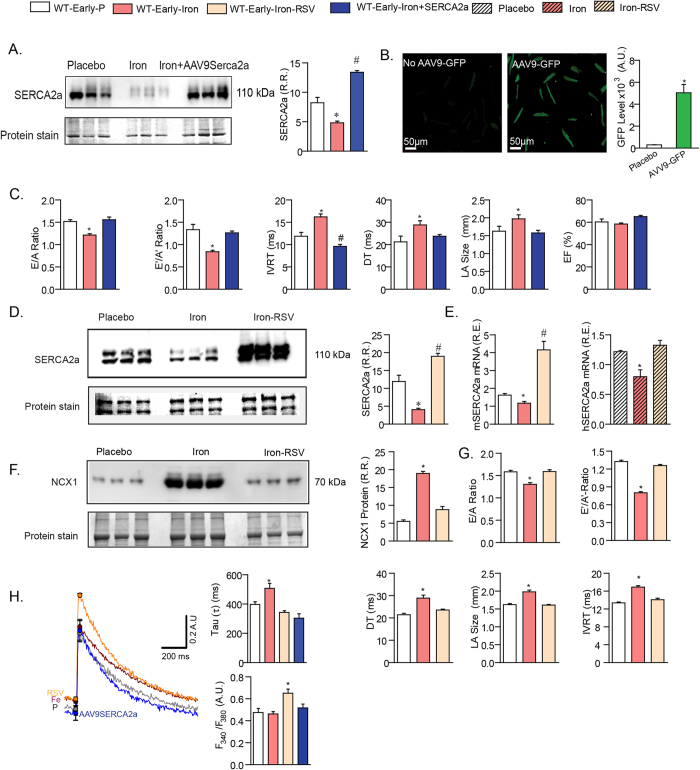
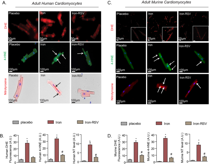
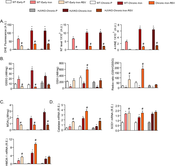
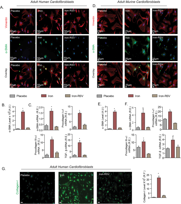
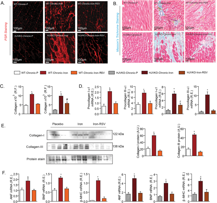
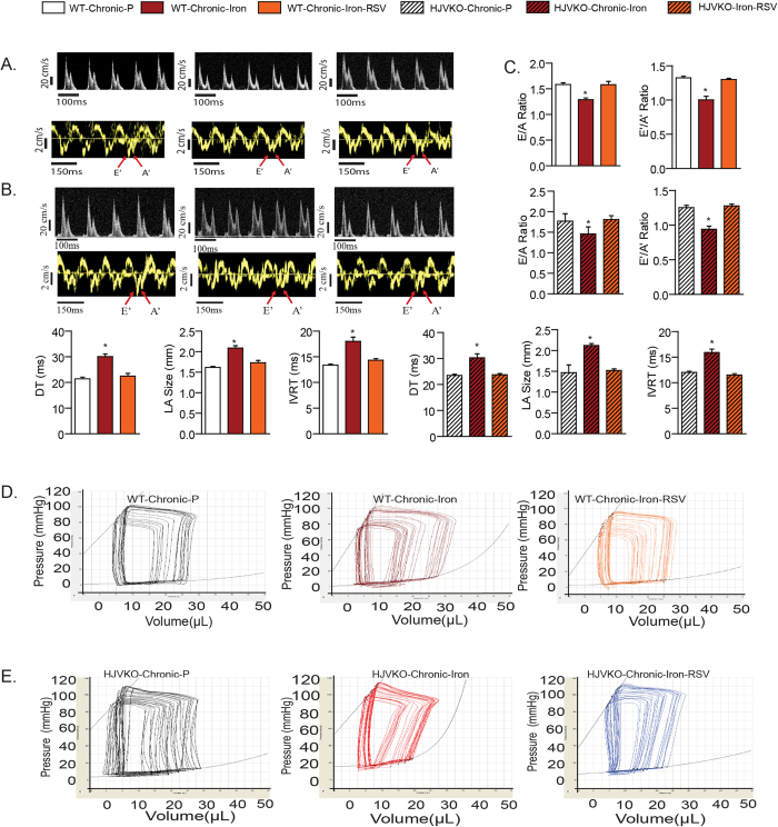
Iron-overload cardiomyopathy is a prevalent cause of heart failure on a world-wide basis and is a major cause of mortality and morbidity in patients with secondary iron-overload and genetic hemochromatosis. We investigated the therapeutic effects of resveratrol in acquired and genetic models of iron-overload cardiomyopathy. Murine iron-overload models showed cardiac iron-overload, increased oxidative stress, altered Ca(2+) homeostasis and myocardial fibrosis resulting in heart disease. Iron-overload increased nuclear and acetylated levels of FOXO1 with corresponding inverse changes in SIRT1 levels in the heart corrected by resveratrol therapy. Resveratrol, reduced the pathological remodeling and improved cardiac function in murine models of acquired and genetic iron-overload at varying stages of iron-overload. Echocardiography and hemodynamic analysis revealed a complete normalization of iron-overload mediated diastolic and systolic dysfunction in response to resveratrol therapy. Myocardial SERCA2a levels were reduced in iron-overloaded hearts and resveratrol therapy restored SERCA2a levels and corrected altered Ca(2+) homeostasis. Iron-mediated pro-oxidant and pro-fibrotic effects in human and murine cardiomyocytes and cardiofibroblasts were suppressed by resveratrol which correlated with reduction in iron-induced myocardial oxidative stress and myocardial fibrosis. Resveratrol represents a clinically and economically feasible therapeutic intervention to reduce the global burden from iron-overload cardiomyopathy at early and chronic stages of iron-overload.


Figures

Figure 1
Figure 1. Iron-overload alters myocardial SIRT1/FOXO1 signaling which is restored by RSV.
(A,B) Western blot analysis and quantification of two major transcriptional factors, Nrf2 and FOXO1, showing no change in Nrf2 levels but increased nuclear levels of FOXO1 in early iron-overloaded hearts (A) with immunofluorescence staining in myocardial tissue with early iron-overload confirming increased nuclear FOXO1 levels as illustrated by the white arrows (B). (C,D) Immunoprecipitated cardiac acetylated FOXO1 increased in response to iron-overload which was markedly suppressed by resveratrol (RSV) with corresponding inverse changes in SIRT1 levels (C) while immunofluorescence staining for FOXO1 (green) and SIRT1 (red) in cultured and stretched murine LV cardiofibroblasts showing that in response iron exposure nuclear FOXO1 increased with reduced SIRT1 levels, while RSV (100 μM) prevents the loss of SIRT1 without affecting the increased total FOXO1 levels (D). Resveratrol therapy increased the phosphorylation of AMPK (threonine-172) in iron-overloaded myocardium (E). SIRT1 activator, SRT1720 (1 μM), prevents iron-induced oxidative stress in cardiomyocytes based on dihydroethidium (DHE) staining for superoxide levels, 4-hydroxynonenal (4-HNE) and nitrotyrosine immunofluorescence (F). R.R. = relative ratio; A.U. = arbitrary unit. n = 3 repeats from n = 2 hearts. *p < 0.05 compared with all other groups; #p < 0.05 compared with the placebo group.
Figure 2
Figure 2. Pathological myocardial remodeling and diastolic dysfunction in early iron-overload is completely rescued by RSV therapy independent of myocardial iron-deposition.
Prussian blue staining and quantification of iron deposition in early iron-overloaded mice showing myocardial iron-overload (A) and altered expression of iron metabolic genes, transferrin receptor 1 (Trfc1), ferroportin (FPN), and ferritin light (L) and heavy (H) chain (B). Resveratrol did not affect myocardial iron deposition or the expression iron metabolism genes (A,B). Echocardiographic assessment of heart function illustrated by transmitral filling pattern (top panel), tissue Doppler (middle panel) and left atrial (LA) size (bottom panel) (C) showing adverse remodeling and diastolic dysfunction in early iron-overloaded wildtype mice. Invasive hemodynamic measurement revealed impaired myocardial relaxation as the primary functional abnormality (D). Resveratrol (RSV) treatment normalized the diastolic dysfunction (C,D) and the expression of myocardial disease markers (E). E’ = early tissue Doppler velocity; A’ = tissue Doppler due to atrial contraction; LA = left atrial; Tau = LV relaxation time constant; dP/dt = rate of change in LV pressure; ANF = atrial natriuretic factor; BNP = brain natriuretic peptide; β-MHC = beta-myosin heavy chain. n = 8 for gene expression analysis; n = 6 for placebo and n = 8 for iron-treated groups. ND = not detected; *p < 0.05 compared with the placebo group.
Figure 3
Figure 3. Early iron-overload cardiomyopathy is driven by downregulation of SERCA2a: rescue with adenoviral transfer of SERCA2a and RSV.
(A,B) Western blot analysis and quantification shows a marked decreased in myocardial SERCA2a level (A) which was prevented by in vivo adeno-viral gene therapy (AAV9) confirmed in isolated adult ventricular cardiomyocytes following in vivo AAV9 delivery of green fluorescent protein (GFP) showing a high yield of efficient gene delivery to the heart (B). Assessment of diastolic function using transthoracic echocardiography showing in vivo gene delivery of SERCA2 normalized the diastolic dysfunction associated with early iron-overload (C). (D,E) Western blot analysis revealed a dramatic corrective action of resveratrol (RSV) on the reduced SERCA2a levels (D) which correlated with the ability of RSV to prevent iron-induced downregulation of Serca2a mRNA expression in mouse (m) and human (h) LV cardiomyocytes (E,F) Western blot analysis of sodium-calcium exchanger-1 (NCX-1) showing increased levels in early iron-overload which was normalized in response to RSV. (G) Functional assessment of heart function showing diastolic dysfunction in early iron-overloaded wildtype mice was completely normalized by RSV therapy. (H) Ca2+ transients in ventricular cardiomyocytes showing elevated diastolic Ca2+ levels and prolongation of Ca2+ decay, and correction by SERCA2a gene therapy and RSV. R.R. = relative ratio; R.E. = relative expression; E = early LV transmitral filling velocity; A = LV transmitral filling due to atrial contraction; DT = deceleration time; LA = left atrial; EF = ejection fraction. E’ = early tissue Doppler velocity; A’ = tissue Doppler due to atrial contraction; IVRT = isovolumetric relaxation time. n = 8–12 for functional studies; n = 8 for expression analysis and n = 3–4 for Western blot analysis. *p < 0.05 compared with all other groups; #p < 0.05 compared with the placebo group.
Figure 4
Figure 4. Iron-induced pro-oxidant effects in human and murine cardiomyocytes and in murine models of iron-overload are prevented by RSV.
(A,B) Isolated adult LV human cardiomyocytes display a pronounced pro-oxidant phenotype after exposure to iron with increased dihydroethidium (DHE) staining for superoxide levels (top), 4-hydroxynonenal (4-HNE) immunofluorescence (middle), nitrotyrosine (NT) immunofluorescence (bottom) (A) and quantification of oxidative stress (B), while resveratrol (RSV; 100 μM) markedly suppressed iron-induced cellular oxidative stress. (C,D) Murine LV cardiomyocytes mirrored similar responses to iron as seen in human LV cardiomyocytes and iron-mediated cellular oxidative stress as illustrated by increased DHE staining, 4-HNE and nitrotyrosine immunofluorescence (C) and quantification of oxidative stress (D) was markedly suppressed by treatment with RSV. DHE fluorescence and is predominantly nuclear while 4-HNE and nitrotyrosine immunofluorescence are more diffuse and highlighted by the white arrows. n = 4 for immunofluorescence analysis; n = 8 for biochemical and gene expression analysis. *p < 0.05 compared with all other groups; #p < 0.05 compared with the placebo group.
Figure 5
Figure 5. Iron-induced oxidative stress in early and chronic murine models of iron-overload is prevented by RSV.
(A) Dihydroethidium fluorescence, 4-hydroxynonenal (4-HNE) and nitrotyrosine (NT) immunostaining confirmed increased myocardial oxidative stress in murine models of iron-overload and the therapeutic effects of resveratrol (RSV). (B) Myocardial levels of reduced glutathione (GSH), oxidized glutathione (GSSG) and the redox ratio, and the myocardial lipid peroxidation product, malondialdehyde (MDA) (C) were altered demonstrating biochemical evidence of increased oxidative damage and reduced anti-oxidant reserve in early and chronic iron-overloaded hearts, markedly corrected by oral RSV therapy. (D) Resveratrol potentiated the upregulation of key anti-oxidant enzymes, catalase (CAT), superoxide dismutase 1 (SOD1) and heme oxygenase 1 (HMOX1), in early and chronic iron-overloaded hearts. A.U. = arbitrary unit; R.E. relative expression; LV = left ventricle; n = 8 for biochemical and expression analyses. *p < 0.05 compared with all other groups; #p < 0.05 compared with the placebo group.
Figure 6
Figure 6. Iron-induced profibrotic effects in human and murine cardiofibroblasts are suppressed by RSV.
(A–C) Cultured and cyclically stretched adult human LV cardiofibroblasts mounted a pro-fibrotic response to exposure to iron (20 μg/ml) resulting in increased immunostaining for alpha-smooth muscle actin (α-SMA) (A,B), and mRNA expression of α-SMA, TGFβ1, pro-collagen type IIIα1, and pro-collagen type Iα1 (C) which was prevented by resveratrol (RSV; 100 μM). (D–F) Murine LV cardiofibroblasts cultured and cyclically stretched showed a similar pro-fibrotic response when exposed to iron (20 μg/ml) with increased levels of alpha-smooth muscle actin (α-SMA) (D,E) and upregulation of the expression of pro-fibrotic genes, pro-collagen Iα1 and IIIα1, α-SMA, TGFβ1, was normalized in response to RSV (100 μM) (F). (G) Human cardiofibroblasts also showed increased collagen I levels in response to iron (20 μg/ml) which was largely prevented by RSV treatment. A.U. = arbitrary unit; R.E. = relative expression; R.F. = relative fraction; α-SMA = alpha smooth muscle actin; TGFβ1 = transforming growth factor beta1; n = 4 for immunofluorescence analysis; n = 8 for expression analysis. *p < 0.05 compared with all other groups.
Figure 7
Figure 7. Increased myocardial fibrosis associated with chronic iron-overload cardiomyopathy is completely rescued by RSV therapy.
(A–C) Histological assessment of myocardial fibrosis using picro-sirius red (PSR) (A) and Masson’s trichrome (B) staining and quantification of fibrosis (C) revealed increased myocardial interstitial and perivascular fibrosis in the chronic iron-overloaded hearts. Expression analysis of myocardial pro-collagen Iα1 and pro-collagen IIIα1 (D) and Western blot analysis of myocardial collagen I and collagen III levels (E) in chronic iron-overloaded hearts revealed increased levels consistent with a pro-fibrotic state. Resveratrol therapy prevented the increased in myocardial fibrosis based on histological, gene expression and Western blot analysis (A–E). Expression analysis of myocardial disease markers in chronic iron-overload models showing a complete normalization of the expression of disease markers in response to resveratrol (RSV) therapy (F). A.U. = arbitrary unit; R.E. = relative expression; R.F. = relative fraction; ANF = atrial natriuretic factor; BNP = brain natriuretic peptide; β-MHC = beta-myosin heavy chain. n = 4 for histological analyses; n = 6 for Western blot and n = 8 for expression analyses. *p < 0.05 compared with all other groups; #p < 0.05 compared with the placebo group.
Figure 8
Figure 8. Resveratrol therapy completely rescued the cardiac dysfunction in chronic iron-overloaded wildtype and hemojuvelin knockout mice.
Echocardiographic assessment of heart function with transmitral filling pattern (top panel) and tissue Doppler (bottom panel) illustrating diastolic dysfunction in chronic iron-overloaded wildtype mice (A) and hemojuvelin knockout (HJVKO) mice (B) and quantification (C) of the echocardiographic assessment showing diastolic dysfunction. Resveratrol (RSV) treatment completely normalized the diastolic dysfunction in wildtype and HJVKO models of chronic iron-overload (A–C). Invasive hemodynamic assessment illustrated by representative pressure-volume tracings confirming load-independent diastolic dysfunction in chronic iron-overloaded wildtype mice (D) and HJVKO mice (E). E = early LV transmitral filling velocity; A = LV transmitral filling due to atrial contraction; E’ = early tissue Doppler velocity; A’ = tissue Doppler due to atrial contraction; DT = deceleration time; LA = left atrial; IVRT = isovolumetric relaxation time. n = 8 for the placebo groups and n = 10–12 for the iron-treated groups. *p < 0.05 compared with all other groups; #p < 0.05 compared with the placebo group.

References

    1. Barton J. C. & Bertoli L. F. Hemochromatosis: the genetic disorder of the twenty-first century. Nat. Med. 2, 394–395 (1996). - PubMed
    1. Andrews N. C. Disorders of iron metabolism. N. Engl. J. Med. 341, 1986–1995 (1999). - PubMed
    1. Pietrangelo A. Hereditary hemochromatosis–a new look at an old disease. N. Engl. J. Med. 350, 2383–2397 (2004). - PubMed
    1. Fleming R. E. & Ponka P. Iron overload in human disease. N. Engl. J. Med. 366, 348–359 (2012). - PubMed
    1. Murphy C. J. & Oudit G. Y. Iron-overload cardiomyopathy: pathophysiology, diagnosis, and treatment. J. Card. Fail. 16, 888–900 (2010). - PubMed

Publication types

MeSH terms