. 2015 Dec 7:6:10065.
doi: 10.1038/ncomms10065.

Corticostriatal pathways contribute to the natural time course of positive mood

Affiliations

Corticostriatal pathways contribute to the natural time course of positive mood

Roee Admon et al. Nat Commun. .

Abstract

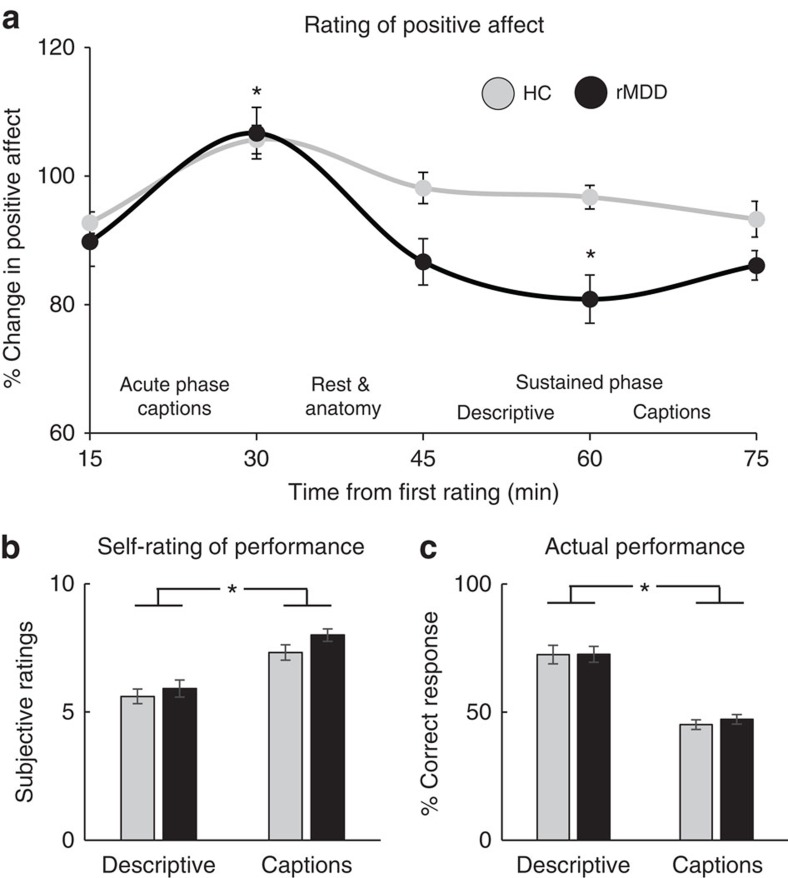
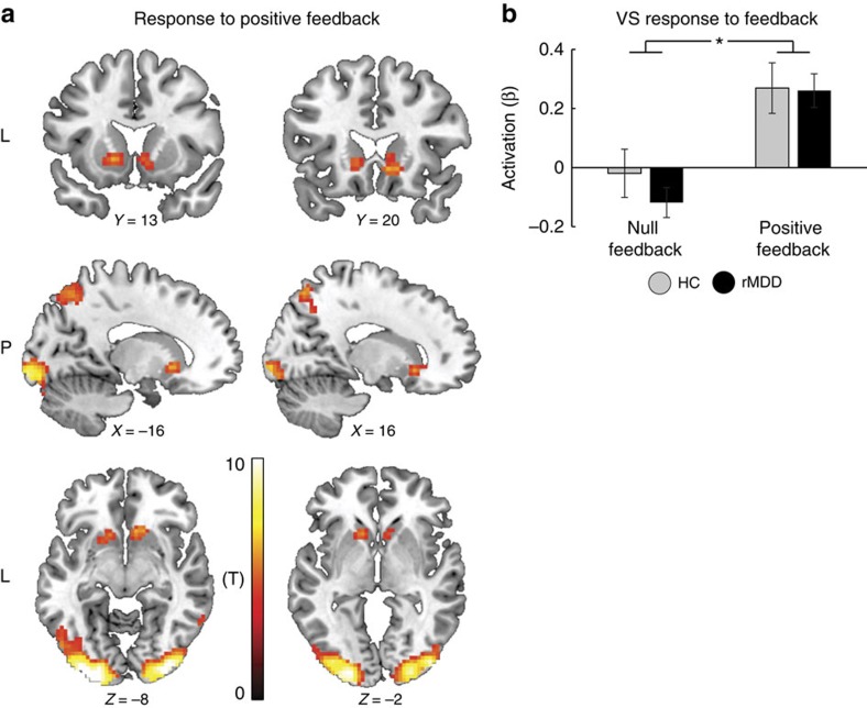
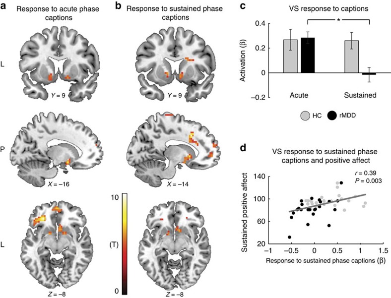
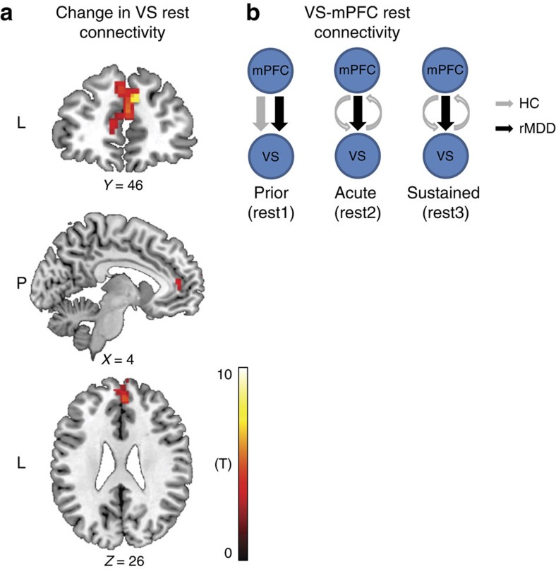
The natural time course of mood includes both acute responses to stimuli and spontaneous fluctuations. To date, neuroimaging studies have focused on either acute affective responses or spontaneous neural fluctuations at rest but no prior study has concurrently probed both components, or how mood disorders might modulate these processes. Here, using fMRI, we capture the acute affective and neural responses to naturalistic positive mood induction, as well as their spontaneous fluctuations during resting states. In both healthy controls and individuals with a history of depression, our manipulation acutely elevates positive mood and ventral striatum activation. Only controls, however, sustain positive mood over time, and this effect is accompanied by the emergence of a reciprocal relationship between the ventral striatum and medial prefrontal cortex during ensuing rest. Findings suggest that corticostriatal pathways contribute to the natural time course of positive mood fluctuations, while disturbances of those neural interactions may characterize mood disorder.


Conflict of interest statement

Over the past 3 years, Dr Pizzagalli has received consulting fees from Otsuka America Pharmaceutical and Pfizer, for activities unrelated to the current research. Dr Admon declares no competing financial interests.

Figures

Figure 1
Figure 1. Task design and timeline.
The task was composed of four phases: The Acute Descriptive phase, in which participants chose which of three suggested sentences best described a cartoon, without receiving any feedback. The Acute Captions Phase, in which participants chose, for each cartoon, which of three suggested humoristic captions won the New Yorker Caption Contest. In this phase, after 14 of their 18 selections, participants saw a screen indicating that they were correct and that they responded faster than average. Following completion of this phase, participants saw a short pre-recorded video in which an actor that presents himself as the director of research is attributing participants' excellent performance in the caption task to their evidently high levels of emotional intelligence and sense of humour (Supplementary Movie 1). Next, the Sustained Descriptive Phase and Sustained Captions Phase were presented, which are identical to the Acute Descriptive and Captions phases, respectively, with the exception that no feedback was given to participants at any point. In addition, three resting state scans were acquired (before the acute phase (rest 1); following the acute phase (rest 2); and following the sustained phase (rest 3)) to investigate the neural basis of spontaneous affect fluctuations in the absence of external stimuli. Finally, participants were asked to rate their current mood state at six time points throughout the task via a visual analogue mood scale (VAMS).
Figure 2
Figure 2. Affective responses to the positive mood induction.
(a) Mixed ANOVA revealed that both groups exhibited a significant increase in positive mood immediately following the mood induction manipulation (that is, following Acute Phase Captions), yet only controls had a sustained elevation in positive affect over time. Specifically, for controls, but not rMDD, positive mood did not decline for at least 30 min following the mood induction. Mixed ANOVA for self-rating of performance in the task (b) and actual performance (c) revealed significant differences between the descriptive and captions tasks, although in opposite directions. Participants, across groups, rated their performance to be better in the captions compared with descriptive task, yet their accuracy was actually better in the descriptive compared with the caption task. Taken together, affective responses suggest that participants were engaged in the task and perceived the positive feedback during the captions task as reliably reflecting their performance, which in turn led to a successful induction of positive mood across groups in an ecologically valid and naturalistic way. Bars ±1 s.e.m. *P<0.05. HC, healthy controls (n=25); rMDD, remitted individuals with a history of recurrent MDD (n=25).
Figure 3
Figure 3. Acute neural responses to the positive feedback.
(a) A whole-brain analysis across groups revealed increased activation in bilateral VS as well as in visual occipital areas in response to the positive feedback compared with null feedback at a significance level adjusted such that Type I error was controlled for all voxels in the brain using the false discovery rate (FDR), with q=0.05 in more than 10 contiguous voxels. (b) Mixed ANOVA revealed that both groups responded to the positive feedback with increased VS activation. Bars ±1 s.e.m. *P<0.05. HC, healthy controls (n=25); rMDD, remitted individuals with a history of recurrent MDD (n=25); VS, ventral striatum.
Figure 4
Figure 4. Neural responses to the captions task.
Whole-brain analyses across groups revealed bilateral VS activation in response to the captions task compared with the descriptive task, at both the acute (a) and sustained (b) phases, at a significance level adjusted such that Type I error was controlled for all voxels in the brain using the false discovery rate (FDR), with q=0.05 in more than 10 contiguous voxels. Additional activations were found in language and semantic brain regions such as inferior frontal gyrus (IFG), superior temporal gyrus (STG), superior frontal gyrus (SFG) and temporal pole. (c) Mixed ANOVA revealed that in the control, but not rMDD group, VS activation in response to the captions task persisted during the sustained phase. For the sake of simplicity, only activations in response to the caption task are presented. (d) Across participants, the level of VS activation in response to the Sustained Phase Captions task was positively correlated with mood rating during the sustained phase. Bars ±1 s.e.m. *P<0.05. HC, healthy controls (n=25); rMDD, remitted individuals with a history of recurrent MDD (n=25); VS, ventral striatum.
Figure 5
Figure 5. Corticostriatal effective connectivity at rest.
(a) A single cluster within the DMN, located in the mPFC (226 voxels; MNI peak coordinates: X=10, Y=50, Z=32; Zscore=4.34; Pcorrected=0.045) exhibited a significant Group by Time (prior, following acute and following sustained) interaction with the VS time course at a significance level adjusted such that Type I error was controlled for all voxels in the DMN using the false discovery rate (FDR), with q=0.05 in more than 10 contiguous voxels. (b) Spectral-DCM analyses revealed that, before the mood induction (rest 1), the best fitting model for both groups was the one describing connectivity from the mPFC to the VS. Following the mood induction (rest 2 and rest 3), controls exhibited a change in their mPFC–VS effective connectivity pattern towards a more reciprocal relation, whereas rMDD maintained their original connectivity pattern throughout. HC, healthy controls (n=25); mPFC, medial prefrontal cortex; rMDD, remitted individuals with a history of recurrent MDD (n=25); VS, ventral striatum.

References

    1. Westermann R., Spies K., Stahl G. & Hesse F. W. Relative effectiveness and validity of mood induction procedures: a meta analysis. Eur. J. Soc. Psychol. 26, 557–580 (1996).
    1. O'Doherty J. P. Reward representations and reward-related learning in the human brain: insights from neuroimaging. Curr. Opin. Neurobiol. 14, 769–776 (2004). - PubMed
    1. Haber S. N. & Knutson B. The reward circuit: linking primate anatomy and human imaging. Neuropsychopharmacology 35, 4–26 (2010). - PMC - PubMed
    1. Laird A. R. et al. Investigating the functional heterogeneity of the default mode network using coordinate-based meta-analytic modeling. J. Neurosci. 29, 14496–14505 (2009). - PMC - PubMed
    1. Andrews-Hanna J. R., Reidler J. S., Sepulcre J., Poulin R. & Buckner R. L. Functional-anatomic fractionation of the brain's default network. Neuron 65, 550–562 (2010). - PMC - PubMed

Publication types

LinkOut - more resources