

Lipid droplets and steroidogenic cells
- PMID: 26639173
- PMCID: PMC4744538
- DOI: 10.1016/j.yexcr.2015.11.024
Lipid droplets and steroidogenic cells
Abstract
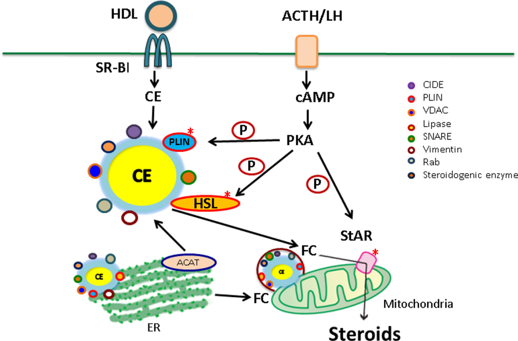
Lipid droplets (LDs) in steroidogenic tissues have a cholesteryl ester (CE) core surrounded by a phospholipid monolayer that is coated with associated proteins. Compared with other tissues, they tend to be smaller in size and more numerous in numbers. These LDs are enriched with PLIN1c, PLIN2 and PLIN3. Both CIDE A and B are found in mouse ovary. Free cholesterol (FC) released upon hormone stimulation from LDs is the preferred source of cholesterol substrate for steroidogenesis, and HSL is the major neutral cholesterol esterase mediating the conversion of CEs to FC. Through the interaction of HSL with vimentin and StAR, FC is translocated to mitochondria for steroid hormone production. Proteomic analyses of LDs isolated from loaded primary ovarian granulosa cells, mouse MLTC-1 Leydig tumor cells and mouse testes revealed LD associated proteins that are actively involved in modulating lipid homeostasis along with a number of steroidogenic enzymes. Microscopy analysis confirmed the localization of many of these proteins to LDs. These studies broaden the role of LDs to include being a platform for functional steroidogenic enzyme activity or as a port for transferring steroidogenic enzymes and/or steroid intermediates, in addition to being a storage depot for CEs.
Keywords: Adrenal gland; Cholesteryl esters; Gonads; Lipid droplet proteins; PLIN; Steroid synthesizing enzymes; Steroidogenesis.
Published by Elsevier Inc.
Figures
References
-
- Murphy DJ. The biogenesis and functions of lipid bodies in animals, plants and microorganisms. Prog Lipid Res. 2001;40(5):325–438. - PubMed
-
- Sato S, et al. Proteomic profiling of lipid droplet proteins in hepatoma cell lines expressing hepatitis C virus core protein. J Biochem. 2006;139(5):921–930. - PubMed
-
- Nestler JETK, Strauss JF., III . Lipoprotein and cholesterol metabolism in cell sthat synthesize steroid hormones. In: Esfahani M, Swaney J, editors. Advances in Cholesterol Research. The Telford Press; 1990. pp. 133–170.
Publication types
MeSH terms
Substances
Grants and funding
LinkOut - more resources
Full Text Sources
Other Literature Sources
Medical
Research Materials
Miscellaneous
