Review
. 2016 Jan;14(1):33-44.
doi: 10.1038/nrmicro.2015.4. Epub 2015 Dec 7.

Mechanistic insights into bacterial AAA+ proteases and protein-remodelling machines

Affiliations
Review

Mechanistic insights into bacterial AAA+ proteases and protein-remodelling machines

Adrian O Olivares et al. Nat Rev Microbiol. 2016 Jan.

Abstract

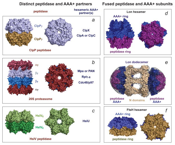
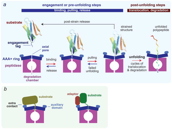
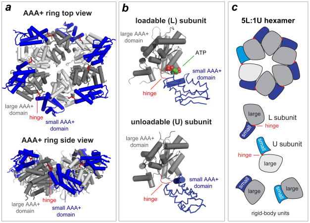
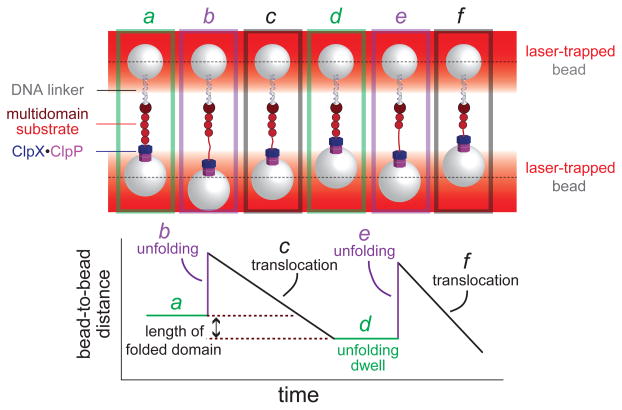
To maintain protein homeostasis, AAA+ proteolytic machines degrade damaged and unneeded proteins in bacteria, archaea and eukaryotes. This process involves the ATP-dependent unfolding of a target protein and its subsequent translocation into a self-compartmentalized proteolytic chamber. Related AAA+ enzymes also disaggregate and remodel proteins. Recent structural and biochemical studies, in combination with direct visualization of unfolding and translocation in single-molecule experiments, have illuminated the molecular mechanisms behind these processes and suggest how remodelling of macromolecular complexes by AAA+ enzymes could occur without global denaturation. In this Review, we discuss the structural and mechanistic features of AAA+ proteases and remodelling machines, focusing on the bacterial ClpXP and ClpX as paradigms. We also consider the potential of these enzymes as antibacterial targets and outline future challenges for the field.


Conflict of interest statement

Competing interests statement

The authors declare no competing interests.

Figures

Figure 1
Figure 1. Self-compartmentalized peptidases are the degradation components of AAA+ proteases
In each panel, a single representative structure is shown. (a) The ClpP peptidase from E. coli (pdb 1TYF) consists of two heptameric rings, uses a Ser–His–Asp catalytic triad for peptide-bond cleavage, and functions with one of three homohexameric AAA+ partners (ClpX, ClpA, or ClpC). In different species, ClpP can consist of 14 identical subunits, distinct homomeric rings or a mixture of subunits in each ring. (b) The 20S proteasome from Thermoplasma acidophilum (pdb 1PMA) has an α7β7β7α7 structure, uses a Thr nucleophile for peptide-bond cleavage, and partners with homohexameric Mpa in bacteria, homohexameric PAN or Cdc48/p97 in archaea, or the heterohexameric Rpt1–6 ring in the eukaryotic 26S proteasome. The α and β rings have seven identical subunits in bacteria and archaea and seven distinct α or β subunits in eukaryotes. (c) The HslV peptidase from Haemophilus influenzae (pdb 1G3I) consists of two homohexameric rings (each subunit is homologous to a β subunit of the 20S proteasome), uses a Thr nucleophile for peptide-bond cleavage, and partners with an HslU homohexamer. (d) The homohexameric Lon protease from Thermococcus onnurineus (pdb 3K1J) is assembled from subunits in which the AAA+ module is fused to the peptidase domain and uses a Ser–Lys dyad for peptide-bond cleavage. (e) Two E. coli Lon hexamers can combine to form a dodecamer, which is stabilized by N-domain interactions that form portals of ~45 Å into the enzyme lumen. The panel shows the E. coli 3LJC and B. subtilis 3M6A structures modeled into a low-resolution electron-density map. (f) The homohexameric Thermotoga maritima FtsH protease (pdb 3KDS) also assembles from subunits in which the AAA+ module is fused to the peptidase domain. FtsH uses an Asp–Zn++ active site for peptide-bond hydrolysis.
Figure 2
Figure 2. Substrate recognition and degradation
(a) Minimal model for recognition, unfolding, translocation and degradation of a single-domain protein by a AAA+ protease. Reaction steps in the forward direction are ATP dependent. In the initial recognition step, a disordered engagement tag in the native protein substrate is bound in the axial pore of the AAA+ ring hexamer. ATP-fuelled conformational changes in the ring then pull on the substrate, which can result either in failed unfolding, substrate release or substrate denaturation. The probability of each of these outcomes depends on substrate stability, as a very stable protein might be bound and released many times resulting in unproductive hydrolysis of a substantial amount of ATP. Following forced unfolding, the denatured polypeptide is processively translocated through the pore and into the peptidase chamber for degradation. (b) Efficient recognition of some protein substrates requires secondary recognition signals, which are substrate sequences that bind to the AAA+ enzyme either directly or via adaptor proteins. In principle, these secondary signals might affect any of the pre-unfolding steps shown in panel a.
Figure 3
Figure 3. ClpX ring structure
(a) Views of the AAA+ ring of E. coli ClpX (pdb 3HWS). In each subunit, the large AAA+ domain is coloured dark or light grey and the small AAA+ domain is coloured purple. Hinges between the large and small domains of each subunit are coloured red. (b) Subunits can adopt either a loadable (L) or an unloadable (U) conformation. In L subunits, ATP binds in a cleft between the large and small AAA+ domains. In U subunits, rotation of the small domain destroys the binding pocket. (c) Cartoon of a 5L:1U ClpX ring showing how six rigid-body units connected by six hinges are created by packing between the small AAA+ domain of a subunit (coloured blue) and the large AAA+ domain of a neighbouring subunit (dark grey for L subunits; light grey for U subunits). Because the ring is topologically closed, changes in the conformation of any single hinge — caused by ATP binding, ATP hydrolysis or product release — propagates around the ring.
Figure 4
Figure 4. Single-molecule force spectroscopy of ClpXP
(a) Cartoon of an optical-trapping experiment. Micron-sized beads, trapped by infrared lasers, are tethered to either ClpXP or a multi-domain substrate via a DNA linker. When ClpXP engages the substrate, ATP-fuelled mechanical activity can be monitored by measuring bead movements relative to the centre of laser focus (dotted lines). (b) ClpXP unfolding of an individual substrate domain (panels a2 and a5) results in an increase in bead-to-bead distance. Translocation of the substrate (panels a3 and a6) results in a decrease in bead-to-bead distance that corresponds to the length of the translocated domain. Periods of no movement are dwells (panels a1 and a4) in which ClpXP tries to unfold the next native domain in the substrate.
Figure 5
Figure 5. Factors influencing the energetic cost of degradation
The average number of ATPs hydrolysed by the ClpXP protease during degradation of a single protein substrate depends upon the protein’s stability and how well it is gripped by ClpX. (a) The axial pore of ClpX contains a loop from each subunit that grips the substrate during protein unfolding. Mutating one or two loops decreases the maximal rate of degradation of a GFP substrate and increases the ATP cost. Thus, maximal unfolding and degradation efficiency requires the combined gripping action of five or six pore loops. (b) Structure of the I27 domain of the human muscle protein titin (pdb 1TIT) and the locations of two mutations (V13P and V15P) that destabilize this domain by removing or disrupting hydrogen bonds. Also shown is an engagement tag at the C-terminus that allows ClpX to recognize and pull on titinI27 variants. The bar graph shows the average ATP cost of ClpXP degradation for the wild-type (WT), V15P and V13P titinI27 domains,. ATP hydrolysed during translocation, terminal unfolding attempts and unfolding attempts that result in substrate release are indicated.
Figure 6
Figure 6. AAA+ remodelling of proteins and protein complexes
(a) A AAA+ remodeling machine breaks a polymer into two pieces by unfolding an interior subunit. Remodelling of the polymer but not the monomer is possible if signals for AAA+ recognition are only properly arranged in the polymer. Related mechanisms may explain severing of microtubules, cell-division rings and amyloid fibers by AAA+ enzymes. (b) Model in which a AAA+ machine enhances the rate of cofactor insertion into a metabolic enzyme by inducing a conformational change in the cofactor binding site. This mechanism appears to be used by mitochondrial ClpX to catalyse incorporation of a cofactor into a haem biosynthesis enzyme. (c) Multi-point binding of a complex to a AAA+ ring, followed by a major conformational change in the ring, might pull the complex apart. This mechanism may account for ClpB remodeling of some bacterial substrates and for disassembly of complexes required for membrane fusion in eukaryotic cells.

References

    1. Oldfield CJ, Dunker AK. Intrinsically disordered proteins and intrinsically disordered protein regions. Annu Rev Biochem. 2014;83:553–584. - PubMed
    1. Sauer RT, Baker TA. AAA+ proteases: ATP-fueled machines of protein destruction. Annu Rev Biochem. 2011;80:587–612. - PubMed
    1. Striebel F, Kress W, Weber-Ban E. Controlled destruction: AAA+ ATPases in protein degradation from bacteria to eukaryotes. Curr Opin Struct Biol. 2009;19:209–217. - PubMed
    1. Matyskiela ME, Martin A. Design principles of a universal protein degradation machine. J Mol Biol. 2013;425:199–213. - PMC - PubMed
    1. Barthelme D, Sauer RT. Identification of the Cdc48•20S proteasome as an ancient AAA+ proteolytic machine. Science. 2012;337:843–846. - PMC - PubMed

Publication types