

Hyalinizing trabecular tumor of the thyroid: diagnosis of a rare tumor using ultrasonography, cytology, and intraoperative frozen sections
- PMID: 26639939
- PMCID: PMC4825213
- DOI: 10.14366/usg.15054
Hyalinizing trabecular tumor of the thyroid: diagnosis of a rare tumor using ultrasonography, cytology, and intraoperative frozen sections
Abstract
Purpose: The goal of this study was to evaluate the clinicopathological and imaging features of thyroid nodules surgically diagnosed as hyaline trabecular tumor (HTT), and to assess the role of cytology and frozen sections (FS) in the diagnosis of HTT.
Methods: This study included 21 thyroid nodules in 21 patients treated from August 2005 to March 2015 (mean age, 53.3 years) who were either diagnosed as HTT or had HTT suggested as a possible diagnosis based on cytology, FS, or the final pathology report. Patients' medical records were retrospectively reviewed for cytopathologic results and outcomes during the course of follow-up. Sonograms were reviewed and categorized.
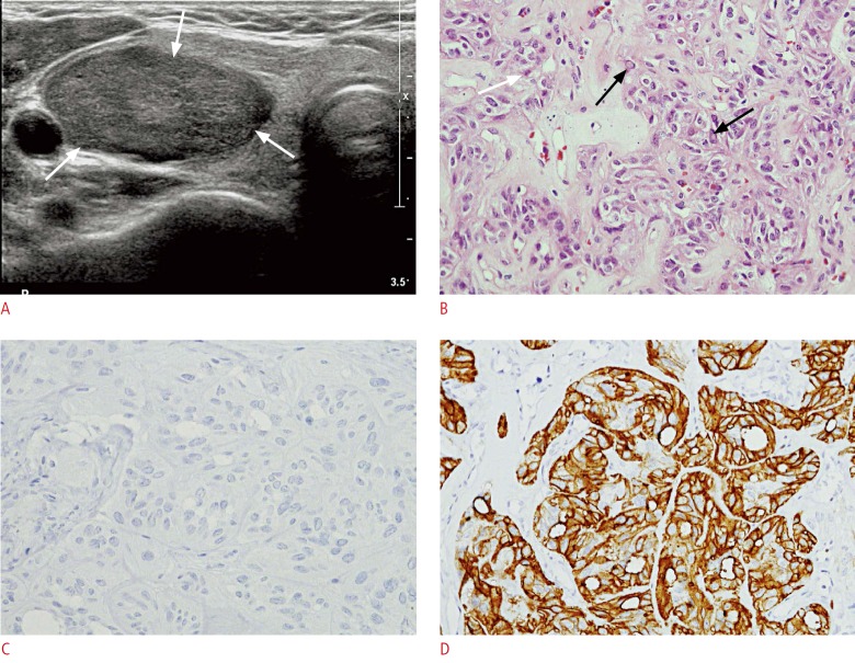
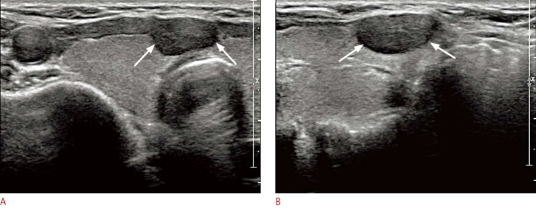
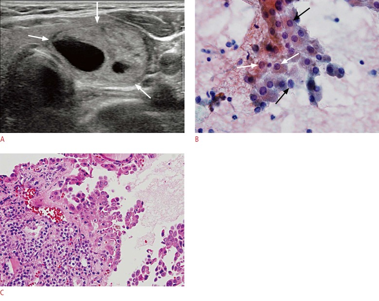
Results: Twelve nodules from 12 patients were surgically confirmed as HTT. Ultrasonography (US)-guided fine needle aspiration (FNA) was performed on 11 nodules, of which six (54.5%) were papillary thyroid carcinoma (PTC) or suspicious for PTC and three (27.3%) were HTT or suspicious for HTT. Intraoperative FS suggested the possibility of HTT in seven nodules, of which four (57.1%) were confirmed as HTT. US-FNA suggested the diagnosis of HTT in 10 nodules, of which three (30.0%) were confirmed as HTT. Common US features of the 12 pathologically confirmed cases of HTT were hypoechogenicity or marked hypoechogenicity (83.4%), absence of calcifications (91.7%), parallel shape (100.0%), presence of vascularity (75.0%), and probable benignity (58.3%).
Conclusion: HTT should be included in the differential diagnosis of solid tumors with hypoechogenicity or marked hypoechogenicity and otherwise benign US features that have been diagnosed as PTC through cytology.
Keywords: Biopsy, fine-needle; Frozen sections; Thyroid gland; Thyroid nodule; Ultrasonography.
Conflict of interest statement
No potential conflict of interest relevant to this article was reported.
Figures
References
-
- Carney JA, Ryan J, Goellner JR. Hyalinizing trabecular adenoma of the thyroid gland. Am J Surg Pathol. 1987;11:583–591. - PubMed
-
- Lee S, Han BK, Ko EY, Oh YL, Choe JH, Shin JH. The ultrasonography features of hyalinizing trabecular tumor of the thyroid are more consistent with its benign behavior than cytology or frozen section readings. Thyroid. 2011;21:253–259. - PubMed
-
- Bondeson L, Bondeson AG. Clue helping to distinguish hyalinizing trabecular adenoma from carcinoma of the thyroid in fine-needle aspirates. Diagn Cytopathol. 1994;10:25–29. - PubMed
-
- Akin MR, Nguyen GK. Fine-needle aspiration biopsy cytology of hyalinizing trabecular adenomas of the thyroid. Diagn Cytopathol. 1999;20:90–94. - PubMed
-
- Evenson A, Mowschenson P, Wang H, Connolly J, Mendrinos S, Parangi S, et al. Hyalinizing trabecular adenoma: an uncommon thyroid tumor frequently misdiagnosed as papillary or medullary thyroid carcinoma. Am J Surg. 2007;193:707–712. - PubMed
LinkOut - more resources
Full Text Sources
Other Literature Sources
