. 2015 Nov 27:9:461.
doi: 10.3389/fncel.2015.00461. eCollection 2015.

Arterial Hypertension Aggravates Innate Immune Responses after Experimental Stroke

Affiliations

Arterial Hypertension Aggravates Innate Immune Responses after Experimental Stroke

Karoline Möller et al. Front Cell Neurosci. .

Abstract

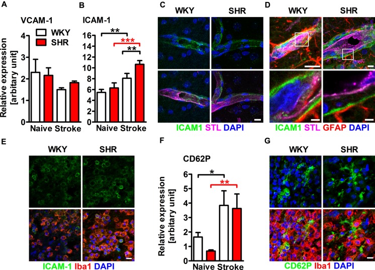
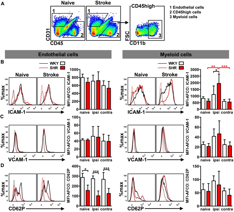
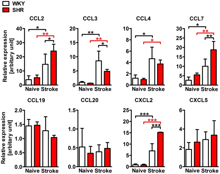
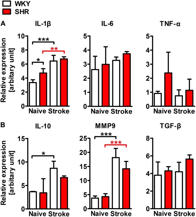
Arterial hypertension is not only the leading risk factor for stroke, but also attributes to impaired recovery and poor outcome. The latter could be explained by hypertensive vascular remodeling that aggravates perfusion deficits and blood-brain barrier disruption. However, besides vascular changes, one could hypothesize that activation of the immune system due to pre-existing hypertension may negatively influence post-stroke inflammation and thus stroke outcome. To test this hypothesis, male adult spontaneously hypertensive rats (SHRs) and normotensive Wistar Kyoto rats (WKYs) were subjected to photothrombotic stroke. One and 3 days after stroke, infarct volume and functional deficits were evaluated by magnetic resonance imaging and behavioral tests. Expression levels of adhesion molecules and chemokines along with the post-stroke inflammatory response were analyzed by flow cytometry, quantitative real-time PCR and immunohistochemistry in rat brains 4 days after stroke. Although comparable at day 1, lesion volumes were significantly larger in SHR at day 3. The infarct volume showed a strong correlation with the amount of CD45 highly positive leukocytes present in the ischemic hemispheres. Functional deficits were comparable between SHR and WKY. Brain endothelial expression of intercellular adhesion molecule 1 (ICAM-1), vascular cell adhesion molecule 1 (VCAM-1), and P-selectin (CD62P) was neither increased by hypertension nor by stroke. However, in SHR, brain infiltrating myeloid leukocytes showed significantly higher surface expression of ICAM-1 which may augment leukocyte transmigration by leukocyte-leukocyte interactions. The expression of chemokines that primarily attract monocytes and granulocytes was significantly increased by stroke and, furthermore, by hypertension. Accordingly, ischemic hemispheres of SHR contain considerably higher numbers of monocytes, macrophages and granulocytes. Exacerbated brain inflammation in SHR may finally be responsible for larger infarct volumes. These findings provide an immunological explanation for the epidemiological observation that existing hypertension negatively affects stroke outcome and mortality.

Keywords: adhesion molecules; animal model; chemokines; hypertension; inflammation; macrophages; neutrophils; stroke.


Figures

FIGURE 1
FIGURE 1
Infarct volume, neurological deficits and myeloid blood cell counts after photothrombotic stroke (PT) in spontaneously hypertensive rats (SHRs) and Wistar Kyoto rats (WKYs). (A) The infarct volume was determined by magnetic resonance (MR) imaging at days 1 and 3 after PT (n = 4/5/8/9). Images show representative series of MR images 3 days after PT in WKY and SHR. (B) Pearson’s correlation of the infarct volume and the amount of CD45 highly positive cells within the ischemic hemisphere (n = 8/9). (C) Neurological deficits were assessed 1 day before, and at days 1 and 3 after PT using the adhesive removal test (ART) asymmetry score (AS) and the modified neurological severity (mNS) score (n = 5/8). (D) Quantification of circulating polymorphonuclear granulocytes (PMN) and monocytes in peripheral blood samples (n = 4/4/5/9). Data are mean values ± standard deviation. p < 0.05, ∗∗p < 0.01.
FIGURE 2
FIGURE 2
Adhesion molecule expression within the ischemic brain of SHRs and WKYs assessed by quantitative real time PCR and immunofluorescence. mRNA expression of (A) vascular cell adhesion molecule 1 (VCAM-1) and (B) intercellular adhesion molecule 1 (ICAM-1) mRNA expression. (C–E) Co-staining of ICAM-1 with Solanum tuberosum lectin (STL), glial fibrillary acidic protein (GFAP) and ionized calcium-binding adapter molecule 1 (Iba1). ICAM-1 is co-expressed by STL+ endothelial cells within the ipsi- and contralateral hemisphere (C), within the parenchymal basement membrane of post-capillary venules adjacent to the ischemic lesion (D) and by Iba1+ myeloid leukocytes within the ischemic lesion (E). mRNA expression of (F) P-selectin (CD62P) and-staining of CD62P and Iba1 (G). Nuclei were counterstained with DAPI; n = 3/3/3/3, data are mean values ± standard deviation. p < 0.05, ∗∗p < 0.01, ∗∗∗p < 0.001. Scale bars: (C) 10 μm; (D) 10 and 1.5 μm; (E) 10 μm; (G) 10 μm.
FIGURE 3
FIGURE 3
Adhesion molecule expression on brain endothelial cells and infiltrated CD45 high leukocytes was determined by flow cytometry. (A) Gating strategy to differentiate CD31+ endothelial cells (1) from CD11b+ myeloid leukocytes (3) in whole hemispheric lysates. Measurement of median fluorescence intensities (MFIs) of (B) intercellular adhesion molecule 1 (ICAM-1), (C) vascular cell adhesion molecule 1 (VCAM-1) and (D) P-selectin (CD62P) on surfaces of endothelial cells and myeloid leukocytes in brains of SHRs and WKYs; n = 3/3/4/6, data are mean values ± standard deviation. p < 0.05, ∗∗p < 0.01, ∗∗∗p < 0.001. AFCO, autofluorescence control; ipsi, ipsilateral; contra, contralateral.
FIGURE 4
FIGURE 4
mRNA expression of the CC chemokine ligands 2 (CCL2), 3 (CCL3), 4 (CCL4), 7 (CCL7) and of the CXC chemokine ligands 2 (CXCL2) and 5 (CXCL5) in whole hemispheric lysates of SHRs and WKYs. n = 3/3/3/3, data are mean values ± standard deviation. p < 0.05, ∗∗p < 0.01, ∗∗∗p < 0.001.
FIGURE 5
FIGURE 5
Differentiation and quantification of leukocyte subsets in the brain of SHRs and WKYs subjected to photothrombotic stroke. (A) Gating strategy to assess CD45 high positive leukocytes, T cells, B cells, polymorphonuclear granulocytes (PMN), monocytes, macrophages and CD11b+ dendritic cells in whole hemispheric lysates. (B) Absolute cell counts of the aforementioned leukocytes subsets were determined by Trucount measurements. (C) The resolution index was calculated by dividing the PMN count through the macrophage count. (D) M1 or M2 macrophage polarization was determined by macrophage expression of CD80 or CD163, respectively; n = 3/3/5/8, data are mean values ± standard deviation. p < 0.05, ∗∗p < 0.01, ∗∗∗p < 0.001. DC, dendritic cells; ipsi, ipsilateral; contra, contralateral.
FIGURE 6
FIGURE 6
mRNA expression of representative M1 (IL-1β, IL-6, and TNF-α) and M2 (IL-10, MMP9, and TGF-β) macrophage markers in whole hemispheric lysates of SHRs and WKYs; n = 3/3/3/3, data are mean values ± standard deviation. p < 0.05, ∗∗p < 0.01, ∗∗∗p < 0.001. Scale bars: (A) 200 μm; (B) 20 μm.

References

    1. Akopov S. E., Simonian N. A., Grigorian G. S. (1996). Dynamics of polymorphonuclear leukocyte accumulation in acute cerebral infarction and their correlation with brain tissue damage. Stroke 27 1739–1743. 10.1161/01.STR.27.10.1739 - DOI - PubMed
    1. Benakis C., Garcia-Bonilla L., Iadecola C., Anrather J. (2014). The role of microglia and myeloid immune cells in acute cerebral ischemia. Front. Cell. Neurosci. 8:461 10.3389/fncel.2014.00461 - DOI - PMC - PubMed
    1. Bratton D. L., Henson P. M. (2011). Neutrophil clearance: when the party is over, clean-up begins. Trends Immunol. 32 350–357. 10.1016/j.it.2011.04.009 - DOI - PMC - PubMed
    1. Buck B. H., Liebeskind D. S., Saver J. L., Bang O. Y., Yun S. W., Starkman S., et al. (2008). Early neutrophilia is associated with volume of ischemic tissue in acute stroke. Stroke 39 355–360. 10.1161/STROKEAHA.107.490128 - DOI - PubMed
    1. Carmichael S. T. (2005). Rodent models of focal stroke: size, mechanism, and purpose. NeuroRx 2 396–409. 10.1602/neurorx.2.3.396 - DOI - PMC - PubMed

LinkOut - more resources