

Mapping abnormal subcortical brain morphometry in an elderly HIV+ cohort
- PMID: 26640768
- PMCID: PMC4625216
- DOI: 10.1016/j.nicl.2015.10.006
Mapping abnormal subcortical brain morphometry in an elderly HIV+ cohort
Abstract
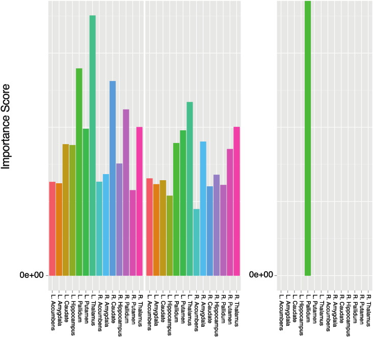
Over 50% of HIV + individuals exhibit neurocognitive impairment and subcortical atrophy, but the profile of brain abnormalities associated with HIV is still poorly understood. Using surface-based shape analyses, we mapped the 3D profile of subcortical morphometry in 63 elderly HIV + participants and 31 uninfected controls. The thalamus, caudate, putamen, pallidum, hippocampus, amygdala, brainstem, accumbens, callosum and ventricles were segmented from high-resolution MRIs. To investigate shape-based morphometry, we analyzed the Jacobian determinant (JD) and radial distances (RD) defined on each region's surfaces. We also investigated effects of nadir CD4 + T-cell counts, viral load, time since diagnosis (TSD) and cognition on subcortical morphology. Lastly, we explored whether HIV + participants were distinguishable from unaffected controls in a machine learning context. All shape and volume features were included in a random forest (RF) model. The model was validated with 2-fold cross-validation. Volumes of HIV + participants' bilateral thalamus, left pallidum, left putamen and callosum were significantly reduced while ventricular spaces were enlarged. Significant shape variation was associated with HIV status, TSD and the Wechsler adult intelligence scale. HIV + people had diffuse atrophy, particularly in the caudate, putamen, hippocampus and thalamus. Unexpectedly, extended TSD was associated with increased thickness of the anterior right pallidum. In the classification of HIV + participants vs. controls, our RF model attained an area under the curve of 72%.
Keywords: CD4, cluster of differentiation; Classification; HIV; JD, Jacobian determinant; MRI; RD, radial distance; Random forest; Shape analysis; Subcortical.
Figures


References
-
- Aylward E.H., Brettschneider P.D., McArthur J.C., Harris G.J., Schlaepfer T.E., Henderer J.D., Barta P.E., Tien A.Y., Pearlson G.D. Magnetic resonance imaging measurement of gray matter volume reductions in HIV dementia. Am. J. Psychiatry. 1995;152:987–994. - PubMed
-
- Aylward E.H., Henderer J.D., McArthur J.C., Brettschneider P.D., Harris G.J., Barta P.E., Pearlson G.D. Reduced basal ganglia volume in HIV-1-associated dementia: results from quantitative neuroimaging. Neurology. 1993;43:2099–2104. - PubMed
Publication types
MeSH terms
Grants and funding
LinkOut - more resources
Full Text Sources
Other Literature Sources
Medical
Research Materials
Miscellaneous
