. 2015 Dec 7;10(12):e0144129.
doi: 10.1371/journal.pone.0144129. eCollection 2015.

TGF-β Controls miR-181/ERK Regulatory Network during Retinal Axon Specification and Growth

Affiliations

TGF-β Controls miR-181/ERK Regulatory Network during Retinal Axon Specification and Growth

Sabrina Carrella et al. PLoS One. .

Abstract

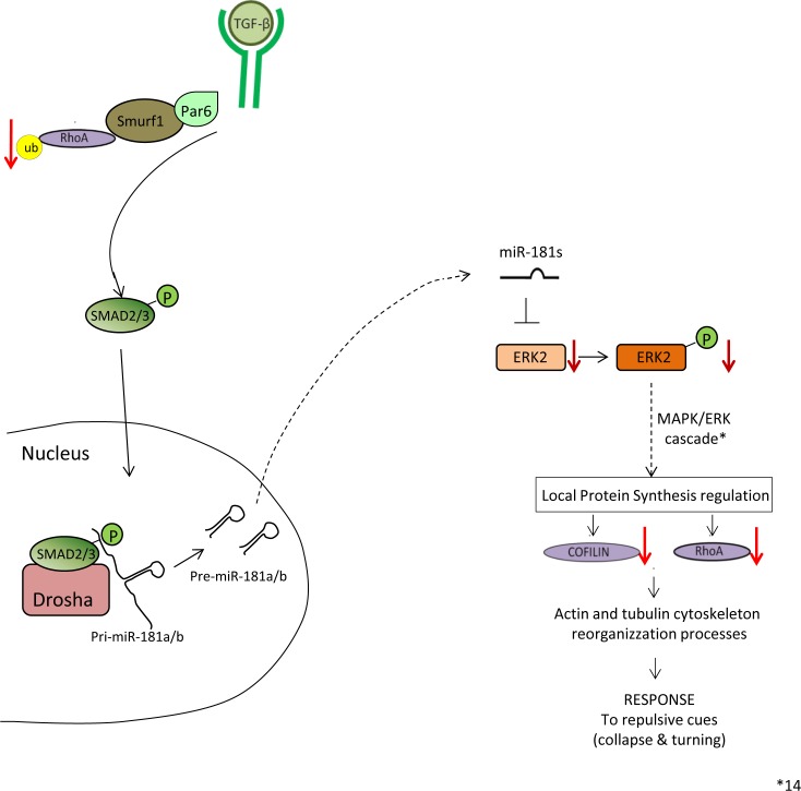
Retinal axon specification and growth are critically sensitive to the dosage of numerous signaling molecules and transcription factors. Subtle variations in the expression levels of key molecules may result in a variety of axonal growth anomalies. miR-181a and miR-181b are two eye-enriched microRNAs whose inactivation in medaka fish leads to alterations of the proper establishment of connectivity and function in the visual system. miR-181a/b are fundamental regulators of MAPK signaling and their role in retinal axon growth and specification is just beginning to be elucidated. Here we demonstrate that miR-181a/b are key nodes in the interplay between TGF-β and MAPK/ERK within the functional pathways that control retinal axon specification and growth. Using a variety of in vivo and in vitro approaches in medaka fish, we demonstrate that TGF-β signaling controls the miR-181/ERK regulatory network, which in turn strengthens the TGF-β-mediated regulation of RhoA degradation. Significantly, these data uncover the role of TGF-β signaling in vivo, for the first time, in defining the correct wiring and assembly of functional retina neural circuits and further highlight miR-181a/b as key factors in axon specification and growth.


Conflict of interest statement

Competing Interests: The authors have declared that no competing interests exist.

Figures

Fig 1
Fig 1. TGF-β pathway down-regulation from early phases of medaka fish embryo development determines alteration of programmed cell death programs in the retina.
(a) MO-tgfβr1 is designed to sterically block the fourth intron-exon splice donor site of the tgfβr1 transcript, causing a partial retention of the intronic sequence as shown by PCR analysis. (b-g) Control (b), MO-tgfβr1 (c), SB43152 (d) and TGF-β- (e) treated control, MO-miR-181a/b (f) and TGF-β treated MO-miR-181a/b (g) medaka fish embryos at stage 32. The MO-tgfβr1 injected embryos showed a phenotype characterized by abnormal body and head structures, including microphthalmia. (h-m) Alteration of the TGF-β pathway from the early stages of development caused an increase of retina cell death as shown by TUNEL assay (h, i). Administration of drugs that lead to a TGF-β pathway down-regulation (j, SB43152) or increase (k, TGF-β) from stage 30 onwards did not cause cell death alteration in control medaka retina. Similarly, no significant alterations were found in MO-miR-181a/b- (l) and TGF-β-treated MO-miR-181a/b (m) retinas. (n) Quantification of TUNEL positive cells; N = 12 eyes were analyzed for each treatment.
Fig 2
Fig 2. TGF-β signaling regulates mature miR-181a/b expression levels.
(a-b) TGF-β pathway inhibition leads to a decrease in the expression levels of mature miR-181a and miR-181b. (a) Administration of SB432542, a TGF-β receptor inhibitor, induced a decrease of miR-181a and miR-181b mature forms in St32 eyes with respect to DMSO treatment, as detected by Taqman assays. The miR-181a and miR-181b reduction was comparable with that observed in the morpholino-mediated inhibition of the TGF-β pathway (MO-tgfβr1) at St32 (b). Data are means +SEM. ***, P <0.001 (t-tests). (c-d) TGF-β treatment (10ng/ml) leads to increased levels of miR-181a and miR-181b mature forms in medaka fish St32 eyes in a transcription-independent manner. (c) Administration of TGF-β (10ng/ml) for 24 h (from St30 to St32) led to the increase of mature miR-181a and miR-181b in St32 eyes, as assessed by Taqman assays. Co-treatment with TFG-β and actinomycin D for 24 h (from St30 to St32) did not alter the TGF-β effect on mature miR-181a/b levels. These results indicate that the TGF-β effect on miR-181a/b expression is not transcription-dependent. Data are means +SEM. ***, P <0.001 (two-way ANOVA). (d) qRT-PCR on RNA extracted from DMSO- and TGF-β-treated St32 medaka fish eyes for all the pri-miR-181a and pri-miR-181b transcripts derived from the different genomic loci present in the medaka fish genome. After 24 h (from St30 to St32) of TGF-β treatment (10ng/ml), there were no significant changes in pri-miR-181a/b levels with respect to DMSO treatment. (e) qRT-PCR on RNA extracted from DMSO-, SB432542- and TGF-β-treated St32 medaka fish eyes. In the SB432542-treated eyes the decrease of miR-181a/b levels led to increased prox1 and erk2 transcript levels, whereas in TGF-β-treated eyes the miR-181a/b increase was accompanied by reduced transcript levels of both prox1 and erk2. Data are means +SEM. **P <0.01; ***, P <0.001 (Two-way ANOVA). (f-g) Representative Western blotting (f) and corresponding quantification (g), showing a decrease of total-, phospho-Erk2 proteins and of its downstream target RhoA in TGF-β-treated St32 medaka fish eyes. Data are means +SEM. **P <0.01 (t-tests).
Fig 3
Fig 3. TGF-β signaling modulates miR-181a/b action in the assembly of retinal circuitry.
(a-e) Retinal frontal sections of St40 DMSO-treated (a), TGF-β-treated (b) control-MO medaka fish embryos, miR-181a/b morphant embryos (c), SB432542-treated embryos (d) and TGF-β-treated (e) miR-181a/b morphant medaka fish embryos processed for Richardson-Romeis staining. Red bars, IPL thickness. Scale bars: 20μm. (f) Quantitative analysis of IPL thickness indicated as the ratio in the central retina between the IPL area and total retinal area. Data are means ± SEM. ***, P <0.001 (one-way ANOVA). (g-j) Representative images of amacrine cells from St40 retinal sections of control-MOs (g), miR-181a/b morphant (h), SB432542-treated control (i) and miR-181a/b morphant treated with TGF-β (j) Six3:eGFP transgenic medaka fish embryos. Cell nuclei are stained with DAPI (blue). GFP (green signal) stains amacrine cell soma and neurites; red arrows, Six3 axon-like structure of amacrine cells; red bars, thickness of the IPL. SB432542 treatment (i) phenocopied the amacrine cell neuritogenesis defects observed in miR-181a/b morphants (h). Addition of TGF-β (from St30 to St40) to miR-181a/b morphants (j) was sufficient to rescue neuritogenesis defects of miR-181a/b morphant transgenic embryos. INL, inner nuclear layer; GCL, ganglion cell layer. Scale bars: 20μm. (k-n) Representative 2-D reconstruction of confocal images of St32 control-MOs (k), miR-181a/b morphant (l), SB432542-treated (m) and TGF-β-treated miR-181a/b morphant (n) Ath5:eGFP transgenic whole-heads. Dotted white lines mark optic nerve routes. Treatment of control-MOs embryos with 80μM SB432542 (m) phenocopied the miR-181a/b-morphant optic nerve length decrease (l). Addition of TGF-β for 24 h (from St30 to St32) to miR-181a/b morphants (n) was sufficient to rescue correct optic nerve growth in Ath5:eGFP morphant embryos. Scale bars: 50μm. OT, optic tectum.
Fig 4
Fig 4. TGF-β administration rescues axon defects in miR-181a/b depleted RGCs.
(a-c) Representative images from primary RGC cultures from St30 control-MOs (a), miR-181a/b morphant (b) and TGF-β-treated miR-181a/b morphant medaka fish embryos (c). The RGC axon length defect was rescued by treatment with TGF-β (c). Scale bars: 10μm. (d) Quantification of RGC axonal length. Data are means +SEM (n = 100) from three independent cell culture experiments. ***P <<0.001 (one-way ANOVA).
Fig 5
Fig 5. TGF-β signaling regulates RhoA levels via two independent and synergistic cascades.
(a) qRT-PCR analysis of erk2 transcripts in total eye RNA derived from St32 control-MOs, miR-181a/b morphants and TGF-β-treated miR-181a/b morphants. The TGF-β-mediated increase of miR-181a/b caused a rescue of miR-181a/b target transcripts, such as prox1 and erk2, in miR-181a/b morphants. (b, c) Representative Western blotting on protein from St32 eyes (b) and corresponding quantification (c) show that administration of TGF-β to MO-miR-181a/b embryos leads to restoration of total-, phospho-Erk2 and RhoA protein levels. When MO-miR-181a/b embryos were treated with both TGF-β and the proteasomal inhibitor MG132, total- and phospho-Erk2 protein levels were still rescued, whereas RhoA levels were only partially rescued. Data are means +SEM.* P <0.05; **P <0.01; *** P <0.001 (two-way ANOVA).
Fig 6
Fig 6. TGF-β signaling regulates erk2 expression by modulating miR-181a/b levels.
(a) qRT-PCR analysis of prox1 and erk2 transcripts in RNA derived from St32 control-MOs, MO-protector-erk2–injected and TGF-β-treated MO-protector-erk2 medaka fish eyes. The TGF-β rescue on the transcript levels of miR-181a/b targets was mediated by the miR-181a/b increase. Indeed this effect on erk2 was completely abolished in the MO-protector-erk2 embryos (a), while other miR-181a/b targets, such as prox1, whose miR-181 binding sites are unaffected by the MO-protector, were still sensitive to TGF-β action. Data are means ± SEM. * P <0.05; ** P <0.01 (two-way ANOVA). (b-d) Retinal frontal sections of St40 Control (b), MO-protector-erk2 (c) and TGF-β-treated MO-protector-erk2 medaka fish embryos (d) processed for Richardson-Romeis staining. Red bars, IPL thickness. Scale bars: 20μm. (e) Quantitative analysis of IPL thickness, indicated as the ratio in the central retina between the IPL area and total retinal area. Data are means ± SEM. *** P <0.0001 (one-way ANOVA). (f-g) Representative Western blotting on protein from St32 eyes (f) and corresponding quantification (g) show that administration of TGF-β to MO-protector-erk2 embryos leads to partial rescue of RhoA protein to levels. When MO-protector-erk2 embryos were treated with both TGF-β and the proteasomal inhibitor MG132, RhoA levels were not rescued anymore. Data are means +SEM. **P <0.05 (one-way ANOVA).
Fig 7
Fig 7. Model of TGF-β cascades in retinal axon specification and growth.
The TGF-β pathway regulates axon growth in the retina via two independent and synergistic pathways: the Par6/Smurf1 and the miR-181/ERK pathways. TGF-β–mediated activation of the Par6/Smurf1 cascade leads to ubiquitination and degradation of RhoA. On the other side, TGF-β also generates increased miR-181a/b levels, enhancing the process of miRNA maturation via activation of the SMAD2/3 protein. In turn, by fine modulation of the MAPK/ERK signaling pathway, miR-181a/b has an inhibitory effect on cofilin and RhoA production.

References

    1. Harada T, Harada C, Parada LF. Molecular regulation of visual system development: more than meets the eye. Genes & development. 2007;21(4):367–78. 10.1101/gad.1504307 . - DOI - PubMed
    1. Duenker N. Transforming growth factor-beta (TGF-beta) and programmed cell death in the vertebrate retina. International review of cytology. 2005;245:17–43. 10.1016/S0074-7696(05)45002-0 . - DOI - PubMed
    1. Close JL, Gumuscu B, Reh TA. Retinal neurons regulate proliferation of postnatal progenitors and Muller glia in the rat retina via TGF beta signaling. Development. 2005;132(13):3015–26. 10.1242/dev.01882 . - DOI - PubMed
    1. Murali D, Kawaguchi-Niida M, Deng CX, Furuta Y. Smad4 is required predominantly in the developmental processes dependent on the BMP branch of the TGF-beta signaling system in the embryonic mouse retina. Investigative ophthalmology & visual science. 2011;52(6):2930–7. 10.1167/iovs.10-5940 - DOI - PMC - PubMed
    1. Massague J, Seoane J, Wotton D. Smad transcription factors. Genes & development. 2005;19(23):2783–810. 10.1101/gad.1350705 . - DOI - PubMed

Publication types

MeSH terms

LinkOut - more resources