. 2016 Jan;19(1):94-101.
doi: 10.1038/nn.4189. Epub 2015 Dec 7.

Injured sensory neuron-derived CSF1 induces microglial proliferation and DAP12-dependent pain

Affiliations

Injured sensory neuron-derived CSF1 induces microglial proliferation and DAP12-dependent pain

Zhonghui Guan et al. Nat Neurosci. 2016 Jan.

Abstract

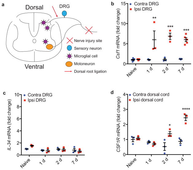
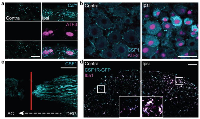
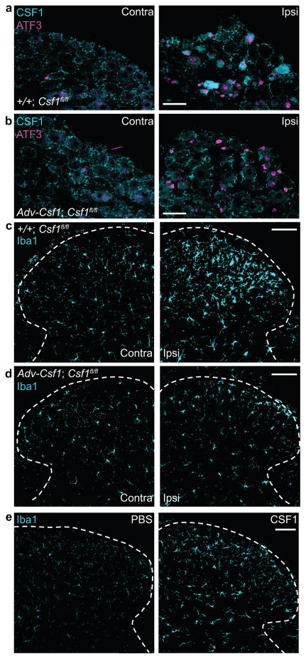
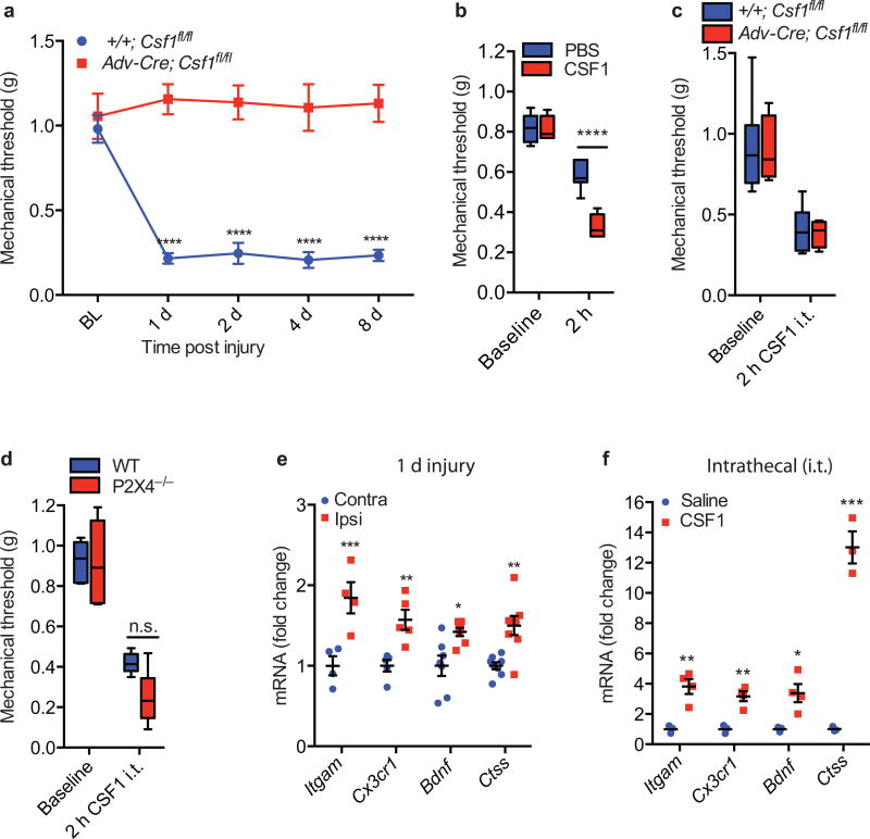
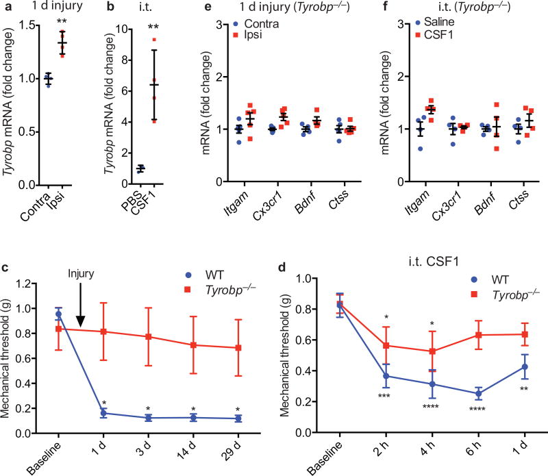
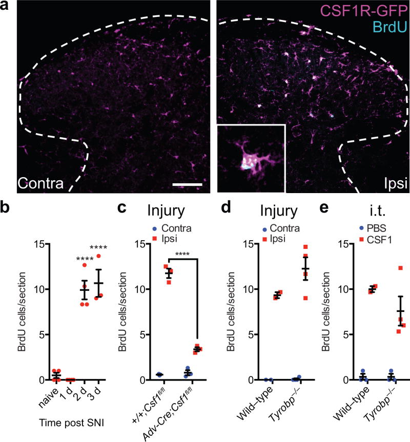
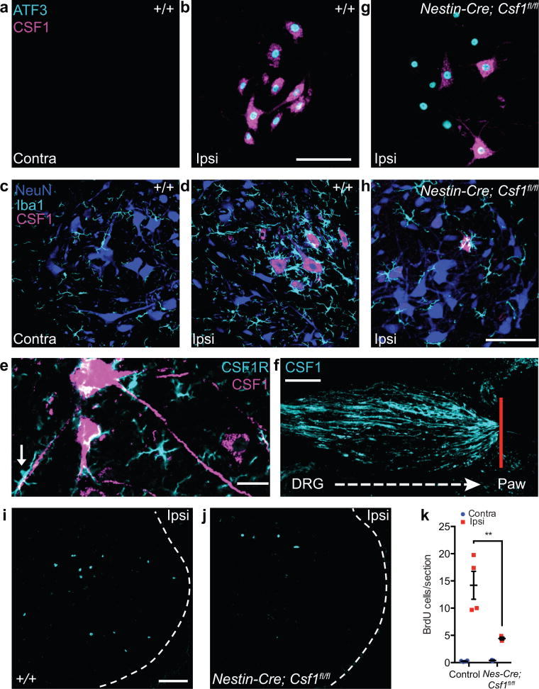
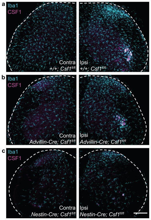
Although microglia have been implicated in nerve injury-induced neuropathic pain, the manner by which injured sensory neurons engage microglia remains unclear. We found that peripheral nerve injury induced de novo expression of colony-stimulating factor 1 (CSF1) in injured sensory neurons. CSF1 was transported to the spinal cord, where it targeted the microglial CSF1 receptor (CSF1R). Cre-mediated sensory neuron deletion of Csf1 completely prevented nerve injury-induced mechanical hypersensitivity and reduced microglial activation and proliferation. In contrast, intrathecal injection of CSF1 induced mechanical hypersensitivity and microglial proliferation. Nerve injury also upregulated CSF1 in motoneurons, where it was required for ventral horn microglial activation and proliferation. Downstream of CSF1R, we found that the microglial membrane adaptor protein DAP12 was required for both nerve injury- and intrathecal CSF1-induced upregulation of pain-related microglial genes and the ensuing pain, but not for microglial proliferation. Thus, both CSF1 and DAP12 are potential targets for the pharmacotherapy of neuropathic pain.


Figures

Figure 1
Figure 1. Csf1 and Csf1r are respectively induced in the DRG and dorsal spinal cord ipsilateral to the peripheral nerve injury
(a) Schematic illustrating relevant neuroanatomy; (b) qRT-PCR illustrates Csf1 induction in the DRG ipsilateral to the peripheral nerve injury, compared to the contralateral side; (c) qRT-PCR shows that there is no induction of IL-34; (d) qRT-PCR illustrates Csf1r induction in the dorsal cord ipsilateral to the nerve injury compared to the contralateral side. N=3 mice/time point.
Figure 2
Figure 2. CSF1 is de novo induced in injured sensory neurons and transported to the spinal cord, where CSF1R is expressed exclusively in microglia
(a) Co-expression of Csf1 mRNA (in situ hybridization) and ATF3 (immunostaining) in injured DRG neurons (1d post injury), compared to contralateral side. Scale bar: 10 μm; (b) Compared to the contralateral side, there is de novo CSF1 (immunostaining) in axotomized, ATF3 positive DRG neurons (1d post injury). Note that there is mild CSF1 immunoreactivity in satellite cells. Scale bar: 50 μm; (c) Concurrent L4 and L5 dorsal root ligation and peripheral nerve injury results in the accumulation of CSF1 protein (immunoreactivity) at the dorsal root ligature (4d post surgery). Red line denotes ligature site (see Fig. 1a). Scale bar: 200 μm; (d) Complete overlap of the microglial markers, Iba1 and GFP in the dorsal horn of a CSF1R-GFP reporter mouse. Both markers increase in the dorsal horn ipsilateral to the nerve injury (3d post injury) compared to the contralateral side. Inset: Control (left) and activated (right) microglia. Note the amoeboid morphology of activated microglia. Scale bar: 100 μm. Inset: maximum projection of Z-stack images.
Figure 3
Figure 3. Sensory neuron-derived CSF1 is necessary and CSF1, by itself, is sufficient for nerve injury-induced microglia activation in the dorsal horn
(a) Injury-induced CSF1 and ATF3 in ipsilateral DRG neurons of a control mouse (+/+; Csf1 fl/fl) (3d post injury). Scale bar: 50 μm; (b) Despite complete loss of CSF1 induction in injured DRG neurons in Adv-Cre;Csf1 fl/fl mice (3d post injury), ATF3 expression persists. Note that the CSF1 immunoreactivity in satellite cells of the DRG is intact in the mutant mice. Scale bar: 50 μm; (c) Peripheral nerve injury-induced microglia activation (increased Iba1 expression) in the ipsilateral dorsal horn (3d post injury) in control animal (+/+; Csf1 fl/fl). Scale bar: 100 μm; (d) Csf1 deletion from sensory neurons (Adv-Cre; Csf1 fl/fl) reduces nerve injury-induced dorsal horn microglia activation. Note that the density and morphology of microglia in the spinal cord contralateral to the nerve injury is comparable between control (+/+; Csf1 fl/fl) and mutant (Adv-Cre; Csf1 fl/fl) mice. Scale bar: 100 μm. (e) Compared to PBS, intrathecal CSF1 activates microglia (increased Iba1 expression) in the dorsal horn. Scale bar: 100 μm. N=3 mice/condition.
Figure 4
Figure 4. Sensory neuron-derived CSF1 is necessary and CSF1, by itself, is sufficient for nerve injury-induced neuropathic pain (mechanical hypersensitivity)
(a) Advillin-Cre mediated Csf1 deletion from sensory neurons prevents the development of nerve injury-induced mechanical hypersensitivity (n=5–6 mice/group); (b) The mechanical hypersensitivity produced by intrathecal CSF1 is significantly greater than that induced by the PBS vehicle (n=7 mice/group) and comparable to that produced by nerve injury; (c) Advillin-Cre mediated Csf1 deletion from sensory neurons had no effect on intrathecal CSF1-induced mechanical hypersensitivity (n=4–6 mice/group); (d) Intrathecal CSF1 induces comparable mechanical hypersensitivity in wild type and P2X4−/− mice (n=6 mice/group); (e) Neuropathic pain-related microglial genes are upregulated in the spinal cord 1 day post injury (n=4 mice/group); (f) Upregulation of the same set of microglial genes occurs in the dorsal horn after intrathecal injection of CSF1 (n=3–4 mice/group). Data are presented as mean ± SEM. n.s. represents “not significant”, with p=0.1332.
Figure 5
Figure 5. DAP12 is required for nerve injury-induced microglia gene upregulation and neuropathic pain (mechanical hypersensitivity)
(a) Upregulation of Tyrobp mRNA (qRT-PCR) in the dorsal cord ipsilateral to nerve injury (1d); (b) Upregulation of Tyrobp mRNA (qRT-PCR) in the spinal cord after intrathecal CSF1; (c) Tyrobp−/− mice do not develop mechanical hypersensitivity after nerve injury (n=5–6 per group); (d) Intrathecal CSF1 does not provoke mechanical hypersensitivity in Tyrobp−/− mice (n=5 per group). The mild hypersensitivity observed in the Tyrobp−/− mice is comparable to that produced by PBS in wild type mice (See Fig. 4b); (e) Tyrobp−/− prevents nerve injury-induced upregulation of neuropathic pain-related microglial genes (1d post injury; n=4–5 mice/group). (f) Tyrobp−/− also prevents intrathecal CSF1-induced microglial gene induction (n=4–5 mice/group).
Figure 6
Figure 6. Sensory neuron-derived CSF1 is necessary and sufficient for nerve injury-induced microglia proliferation in the dorsal horn
(a) Double-labeling for BrdU (to label proliferating microglia) and GFP in the dorsal horn of a CSF1R-GFP mouse (SNI 3d). Note that all the BrdU+ cells express CSF1R. Inset: BrdU incorporation is limited to CSF1R-expressing microglia. Scale bar: 100 μm; Inset: maximum projection of Z-stack images. (b) Microglia proliferation (BrdU incorporation) begins 2d after injury (n=3–4 mice/group); (c) Advillin-Cre-mediated deletion of Csf1 from sensory neurons significantly decreases injury-induced dorsal horn microglia proliferation (3d post injury, n=3 mice/group); (d) Comparable nerve injury-induced microglia proliferation in wild type and Tyrobp−/− mice (3d post injury, n=3–4 mice/group); (e) Intrathecal CSF1 induces dorsal horn microglia proliferation in wild-type and this proliferation is preserved in Tyrobp−/− mice (n=3–4 mice/group, 3d post injury). Data are presented as mean ± SEM.
Figure 7
Figure 7. CSF1 is upregulated in injured motoneurons and is required for nerve injury-induced microglia activation and proliferation in the spinal cord ventral horn
(a) Neither CSF1 nor ATF3 was expressed in ventral horn motoneurons contralateral to nerve injury (3 d post injury). Scale bar represents 100 μm for (a), (b) and (g). (b) Coexpression of CSF1 and ATF3 (immunostaining) in axotomized ventral horn motoneurons (3 d post injury). (c) No microglia activation in contralateral ventral horn (8 d post injury). Scale bar represents 100 μm for (c), (d), and (h). (d) Activated ventral horn microglia (enhanced Iba1 expression) surrounded CSF1-expressing motoneurons (8 d post injury). (e) Motoneuron axons transported CSF1. Note the apposition of CSF1R-expressing microglia and a CSF1+ motoneuron dendrite (arrow). Scale bar represents 25 μm. (f) CSF1 accumulation at the peripheral nerve injury site (4 d post injury). Red line denotes ligature site. Scale bar represents 100 μm, (g) CSF1 upregulation was significantly reduced in Nestin–Cre; Csf1fl/fl mice, despite the persistent motoneuronal ATF3 expression. Given that ~30% of motoneurons continued to express CSF1 after injury, Nestin–Cre was likely not expressed in all motoneurons. (h) Csf1 deletion from the majority of CNS neurons (Nestin–Cre; Csf1fl/fl) reduced ventral horn microglia activation after injury. (i,j) Peripheral nerve injury (3 d) induced microglia proliferation in the ventral horn in wild–type (i), and this was greatly attenuated when Csf1 was deleted from CNS neurons (Nestin–Cre; Csf1fl/fl) (j). Scale bar represents 100 μm for (i) and (j). (k) Quantification of i and j (n = 3–4 mice per group). **P ≤ 0.01.
Figure 8
Figure 8. Cre-mediated neuronal Csf1 deletion reveals topographic distribution of microglia activation after nerve injury
(a) Peripheral nerve injury-induced microglia activation (increased Iba1 expression) in the ipsilateral dorsal and ventral horn, and upregulation of CSF1 in ventral horn motoneurons (3d post injury) in a control animal (+/+; Csf1 fl/fl); (b) Csf1 deletion from sensory neurons (Adv-Cre; Csf1 fl/fl) reduces injury-induced dorsal horn microglia activation. There is no effect on ventral horn microglia activation or on motoneuronal CSF1 induction; (c) Csf1 deletion from the majority of CNS neurons (Nestin-Cre; Csf1 fl/fl) reduces ventral horn microglia activation after injury (See also Fig. 7h). Note that the density of microglia in the spinal cord contralateral to the nerve injury is reduced in the mutant mice. Despite this overall reduction, microglia are still activated in the dorsal horn ipsilateral to the nerve injury in the mutant mice. Scale bar: 200 μm.

Comment in

References

    1. Basbaum AI, Bautista DM, Scherrer G, Julius D. Cellular and molecular mechanisms of pain. Cell. 2009;139:267–284. - PMC - PubMed
    1. von Hehn CA, Baron R, Woolf CJ. Deconstructing the neuropathic pain phenotype to reveal neural mechanisms. Neuron. 2012;73:638–652. - PMC - PubMed
    1. Backonja M, Woolf CJ. Future directions in neuropathic pain therapy: closing the translational loop. Oncologist. 2010;15(Suppl 2):24–29. - PubMed
    1. Sieweke MH, Allen JE. Beyond stem cells: self-renewal of differentiated macrophages. Science. 2013;342:1242974. - PubMed
    1. Salter MW, Beggs S. Sublime microglia: expanding roles for the guardians of the CNS. Cell. 2014;158:15–24. - PubMed

Publication types

MeSH terms

Substances