. 2016 Jan;48(1):74-8.
doi: 10.1038/ng.3465. Epub 2015 Dec 7.

A missense mutation in TFRC, encoding transferrin receptor 1, causes combined immunodeficiency

Affiliations

A missense mutation in TFRC, encoding transferrin receptor 1, causes combined immunodeficiency

Haifa H Jabara et al. Nat Genet. 2016 Jan.

Abstract

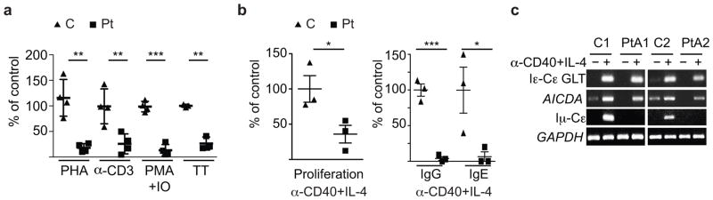
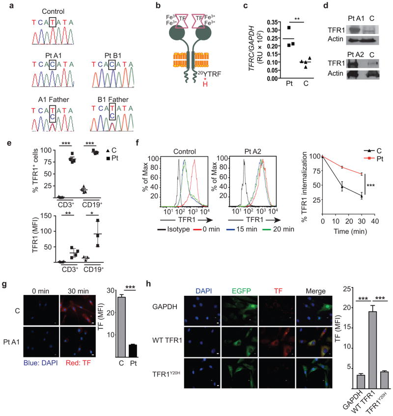
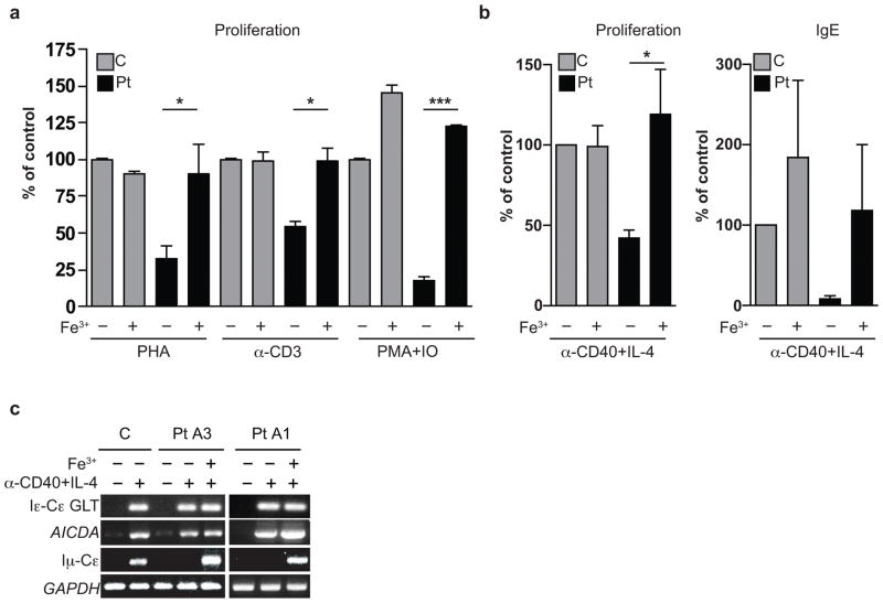
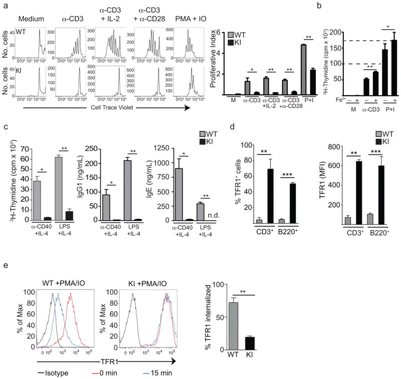
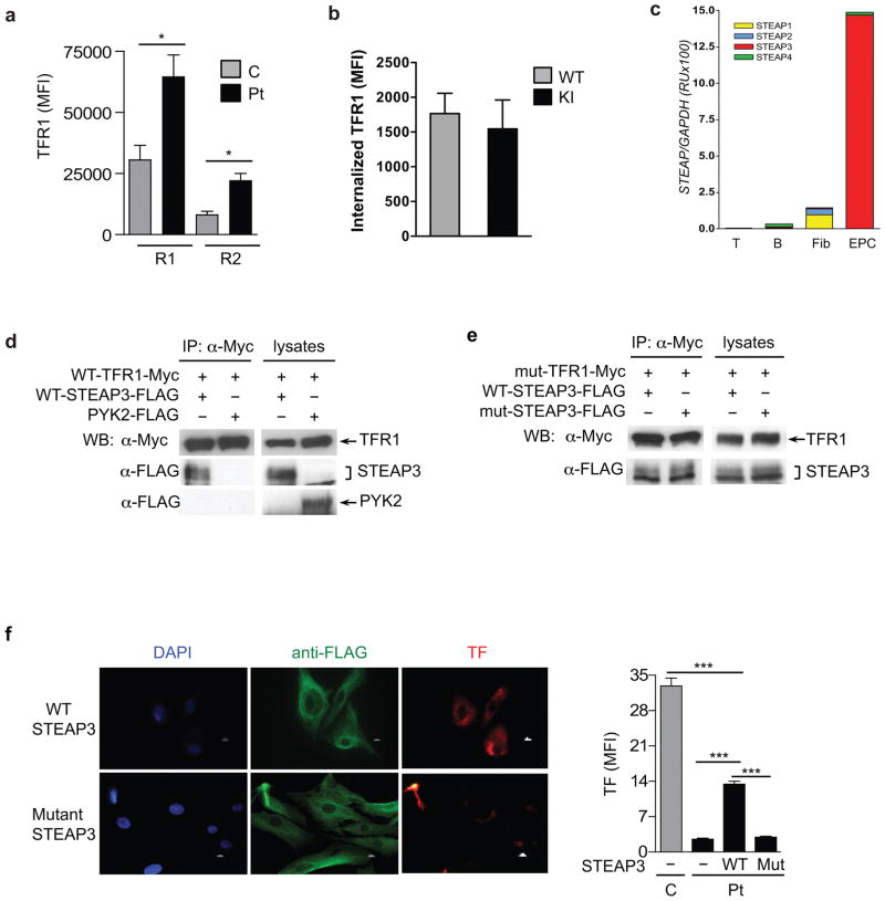
Patients with a combined immunodeficiency characterized by normal numbers but impaired function of T and B cells had a homozygous p.Tyr20His substitution in transferrin receptor 1 (TfR1), encoded by TFRC. The substitution disrupts the TfR1 internalization motif, resulting in defective receptor endocytosis and markedly increased TfR1 expression on the cell surface. Iron citrate rescued the lymphocyte defects, and expression of wild-type but not mutant TfR1 rescued impaired transferrin uptake in patient-derived fibroblasts. Tfrc(Y20H/Y20H) mice recapitulated the immunological defects of patients. Despite the critical role of TfR1 in erythrocyte development and function, patients had only mild anemia and only slightly increased TfR1 expression in erythroid precursors. We show that STEAP3, a metalloreductase expressed in erythroblasts, associates with TfR1 and partially rescues transferrin uptake in patient-derived fibroblasts, suggesting that STEAP3 may provide an accessory TfR1 endocytosis signal that spares patients from severe anemia. These findings demonstrate the importance of TfR1 in adaptive immunity.


Figures

Figure 1
Figure 1. Lymphocyte dysfunction in Patients A1–A3 and B1
(a) PBMC proliferation to phytohemagglutinin (PHA), anti-CD3 antibody (α-CD3), phorbol 12-myristate 13-acetate and ionomycin (PMA+IO), and tetanus toxoid antigen (TT). There were insufficient cells from Patient B1 for proliferation studies to TT. (b) B cell proliferation and IgG and IgE production to anti-CD40+IL-4 stimulation, and (c) molecular events in IgE switching as shown by PCR of B cell cDNA. GLT, germline transcript; AICDA, activation induced cytidine deaminase; GAPDH, glyceraldehyde 3-phosphate dehydrogenase used as a house-keeping gene control. Patient B1 was not studied in these assays because of his lack of circulating B cells. Symbols represent individual patients (Pt) and controls (C). Graphs display means ± SEM; *P<0.05, **P<0.01, ***P<0.001.
Figure 2
Figure 2. TFRC mutation, increased TfR1 surface expression, and impaired internalization of mutant TfR1 protein
(a) Chromatograms depicting the c.58T>C TFRC mutation. (b) Schematic representation of the TfR1 dimer and the p.Tyr20His mutation within the internalization motif. (c) TFRC mRNA levels from Patients A1-A3, normalized to GAPDH. (d) TfR1 protein expression in PBMCs, with actin as a loading control. (e) Pooled FACS data of TfR1 expression on CD3+ T cells from Patients A1–3 and B1 and on CD19+ B cells from Patients A13, compared to controls. TfR1 expression on CD19+ B cells was not studied in Patient B1 due to his lack of circulating B cells after receiving anti-CD20 monoclonal antibody. MFI, mean fluorescence intensity. (f) Representative and pooled TfR1 internalization data after TfR1 crosslinking in EBV-transformed B cells from Patients A1–3 and controls. (g,h) Immunofluorescence analysis of Alexa 568-labeled transferrin (TF) uptake 30 minutes after TF addition by (g) serum-starved fibroblasts and (h) transduced patient fibroblasts, with the graphs in (g) and (h) showing the pooled results from Patients A1A3. Scale bar: 10 μm. Results represent net MFI after background subtraction. Graphs display means ± SEM (n=2–3/group). Symbols represent individual patients as indicated with means ± SEM; *P<0.05, **P<0.01, ***P<0.001.
Figure 3
Figure 3. Correction of lymphocyte defects in Patients A1–3 with iron citrate
Effect of addition of iron citrate on: (a) T cell proliferation to three stimuli, (b) B cell proliferation and IgE synthesis to anti-CD40+IL-4, and (c) molecular events in IgE isotype switching. Bars represent means ± SEM from three independent experiments; *P<0.05, ***P<0.001.
Figure 4
Figure 4. Lymphocyte defects and impaired TfR1 internalization in TfrcY20H/Y20H mice
(a) Representative T cell proliferation measured by CellTrace Violet dye dilution and pooled data from three mice studied in three independent experiments. (b) Effect of addition of iron citrate on T cell proliferation in three mice studied in three independent experiments. M, medium; P+I, PMA and ionomycin. Dashed lines represent the normal values for T cell proliferation to anti-CD3 stimulation and PMA and ionomycin. (c) B cell proliferation and IgG1 and IgE secretion following stimulation from three mice of each genotype in two independent experiments; n.d., not detected. (d) Surface TfR1 expression, determined by FACS analysis, on unstimulated splenic T and B cells, representing five mice of each genotype in five independent experiments. (e) Representative and pooled FACS analysis of TfR1 expression on PMA and ionomycin-activated T cells after TfR1 crosslinking in three TfrcY20H/Y20H (KI) and three wild type (WT) mice in three independent experiments. Error bars represent means ± SEM. *P<0.05, **P<0.01, ***P<0.001.
Figure 5
Figure 5. Partial rescue of defective transferrin uptake in patient fibroblasts by STEAP3 expression
(a) TfR1 surface expression on glycophorin A (CD235a)+ erythroid precursor cells (EPCs) for early (R1) and intermediate (R2) normoblasts, from Patients A1 and A3 and three controls. (b) Internalization of TfR1 after 30 minutes of TfR1 crosslinking on EPCs from wild type (WT) and TfrcY20H/Y20H (KI) mice. (c) mRNA expression of STEAP genes in cells from three controls. Fib, fibroblasts; EPC, erythroid precursor cells. (d) Co-immunoprecipitation (IP) and western blotting (WB) of Myc-tagged wild type (WT) human TfR1 and FLAG-tagged WT murine STEAP3 or PYK2 (as negative control) in co-transfected in HEK293T cells. Immunoblotting of lysates without IP served as a positive control. (e) Co-immunoprecipitation and western blotting of Myc-tagged mutant TfR1Y20H (mut-TfR1-Myc) and FLAG-tagged WT or mutant STEAP3Y288H (mut-STEAP3-FLAG) co-transfected in HEK293T cells. (f) Alexa 568-labeled transferrin (TF) uptake by patient fibroblasts transfected with WT or mutant STEAP3Y288H, and quantitation of uptake compared to untransfected patient and control fibroblasts, assayed in parallel (n=3 per group). Scale bar: 10 μm. Mut, mutant STEAP3Y288H. Error bars represent means ± SEM; *P<0.05, ***P<0.001.

Comment in

References

    1. Al-Herz W, Bousfiha A, Casanova J–L, Chatila T, et al. Front Immunol. 2014;5:1–133. - PMC - PubMed
    1. Jabara HH, Fu SM, Geha RS, Vercelli D. CD40 and IgE: synergism between anti-CD40 monoclonal antibody and interleukin 4 in the induction of IgE synthesis by highly purified human B cells. J Exp Med. 1990;172:1861–4. - PMC - PubMed
    1. McHeyzer-Williams LJ, McHeyzer-Williams MG. Antigen-specific memory B cell development. Annual Review of Immunology. 2005;23:487–513. - PubMed
    1. Jing SQ, Spencer T, Miller K, Hopkins C, Trowbridge IS. Role of the human transferrin receptor cytoplasmic domain in endocytosis: localization of a specific signal sequence for internalization. J Cell Biol. 1990;110:283–94. - PMC - PubMed
    1. Andrews NC. Iron homeostasis: insights from genetics and animal models. Nat Rev Genet. 2000;1:208–17. - PubMed

Publication types

MeSH terms