. 2015 Dec 7;10(12):e0144342.
doi: 10.1371/journal.pone.0144342. eCollection 2015.

Validity of Estimation of Pelvic Floor Muscle Activity from Transperineal Ultrasound Imaging in Men

Affiliations

Validity of Estimation of Pelvic Floor Muscle Activity from Transperineal Ultrasound Imaging in Men

Ryan E Stafford et al. PLoS One. .

Abstract

Purpose: To investigate the relationship between displacement of pelvic floor landmarks observed with transperineal ultrasound imaging and electromyography of the muscles hypothesised to cause the displacements.

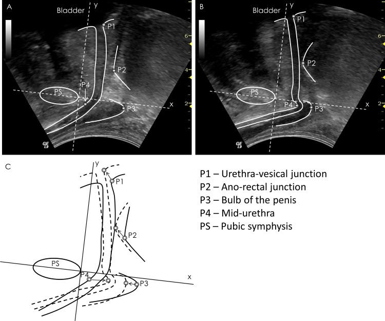
Materials and methods: Three healthy men participated in this study, which included ultrasound imaging of the mid-urethra, urethra-vesical junction, ano-rectal junction and bulb of the penis. Fine-wire electromyography electrodes were inserted into the puborectalis and bulbocavernosus muscles and a transurethral catheter electrode recorded striated urethral sphincter electromyography. A nasogastric sensor recorded intra-abdominal pressure. Tasks included submaximal and maximal voluntary contractions, and Valsalva. The relationship between each of the parameters measured from ultrasound images and electromyography or intra-abdominal pressure amplitudes was described with nonlinear regression.

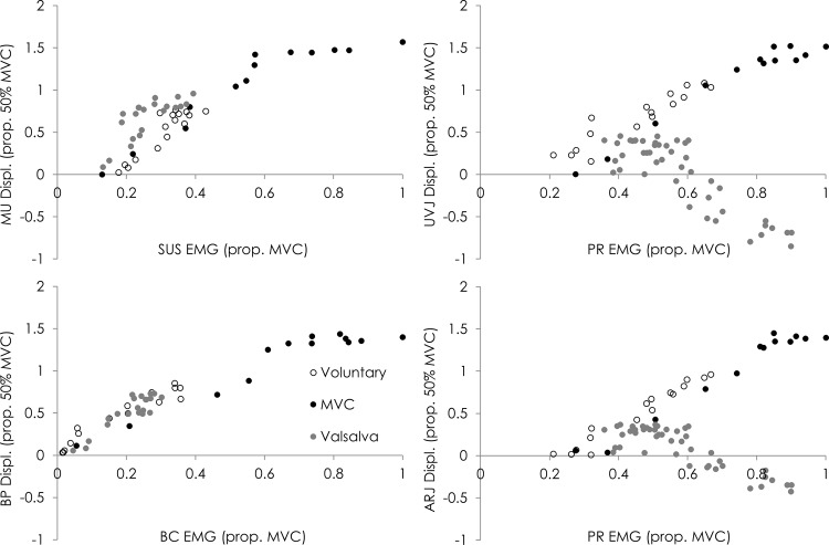
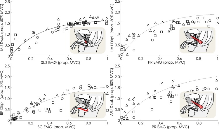
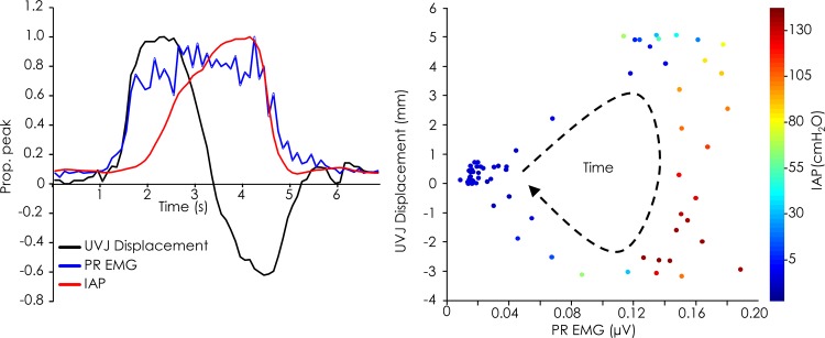
Results: Strong, non-linear relationships were calculated for each predicted landmark/muscle pair for submaximal contractions (R2-0.87-0.95). The relationships between mid-urethral displacement and striated urethral sphincter electromyography, and bulb of the penis displacement and bulbocavernosus electromyography were strong during maximal contractions (R2-0.74-0.88). Increased intra-abdominal pressure prevented shortening of puborectalis, which resulted in weak relationships between electromyography and anorectal and urethravesical junction displacement during all tasks.

Conclusions: Displacement of landmarks in transperineal ultrasound imaging provides meaningful measures of activation of individual pelvic floor muscles in men during voluntary contractions. This method may aid assessment of muscle function or feedback for training.


Conflict of interest statement

Competing Interests: The authors have declared that no competing interests exist.

Figures

Fig 1
Fig 1. Method for calculation of pelvic floor displacements.
Representative transperineal ultrasound images recorded in the (A) relaxed and (B) contracted states with pelvic structure borders and points of interest superimposed on the images. (C) Combined borders from the two images to indicate change in position. The circles indicate the points of interest used for displacement calculations, and the arrows indicate direction of displacement with voluntary contraction.
Fig 2
Fig 2. Data of the relationships between landmark displacement and muscle activity for a representative participant for each task; sub-maximal voluntary contraction, maximal voluntary contraction (MVC) and Valsalva.
MU–mid-urethra, UVJ–urethra-vesical junction, ARJ–ano-rectal junction, BP–bulb of penis, SUS–striated urethral sphincter, PR–puborectalis, BC–bulbocavernosus.
Fig 3
Fig 3. Individual (shapes) and group data (lines) of the relationships between displacement of pelvic floor landmarks observed with transperineal US imaging and activity of the muscles predicted to cause the displacement.
Lines are generated using the mean R2, asymptote and curvature for the group. MU–mid-urethra, UVJ–urethra-vesical junction, ARJ–ano-rectal junction, BP–bulb of penis, SUS–striated urethral sphincter, PR–puborectalis, BC–bulbocavernosus.
Fig 4
Fig 4. The relationship between displacement at the urethra-vesical junction (UVJ), puborectalis (PR) EMG and intra-abdominal pressure (IAP) for an individual participant during a repetition of the MVC task.
Data normalised to peak are shown (left) in addition to the corresponding raw data (right).

References

    1. Davis SN, Morin M, Binik YM, Khalife S, Carrier S. Use of pelvic floor ultrasound to assess pelvic floor muscle function in Urological Chronic Pelvic Pain Syndrome in men. J Sex Med. 2011;8:3173–80. 10.1111/j.1743-6109.2011.02452.x - DOI - PubMed
    1. Geraerts I, Van Poppel H, Devoogdt N, Van Cleynenbreugel B, Joniau S, Van Kampen M. Prospective evaluation of urinary incontinence, voiding symptoms and quality of life after open and robot-assisted radical prostatectomy. BJU Int. 2013;112:936–43. 10.1111/bju.12258 - DOI - PubMed
    1. Nahon I, Waddington G, Adams R, Dorey G. Assessing muscle function of the male pelvic floor using real time ultrasound. Neurourol Urodyn. 2011;30:1329–32. 10.1002/nau.21069 - DOI - PubMed
    1. Stafford RE, Ashton-Miller JA, Constantinou CE, Hodges PW. Novel insight into the dynamics of male pelvic floor contractions through transperineal ultrasound imaging. J Urol. 2012;188:1224–30. 10.1016/j.juro.2012.06.028 - DOI - PMC - PubMed
    1. Stafford RE, Ashton-Miller JA, Constantinou CE, Hodges PW. A New Method to Quantify Male Pelvic Floor Displacement From 2D Transperineal Ultrasound Images. Urology. 2013;81:685–9. 10.1016/j.urology.2012.11.034 - DOI - PMC - PubMed

Publication types