. 2016 Mar;17(3):250-8.
doi: 10.1038/ni.3333. Epub 2015 Dec 7.

NLRP3 activation and mitosis are mutually exclusive events coordinated by NEK7, a new inflammasome component

Affiliations

NLRP3 activation and mitosis are mutually exclusive events coordinated by NEK7, a new inflammasome component

Hexin Shi et al. Nat Immunol. 2016 Mar.

Abstract

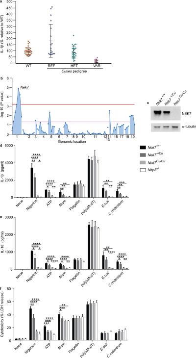
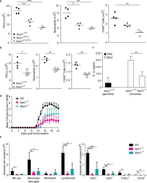
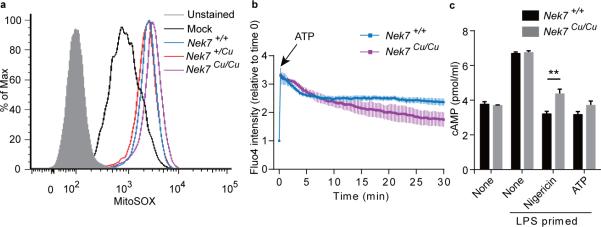
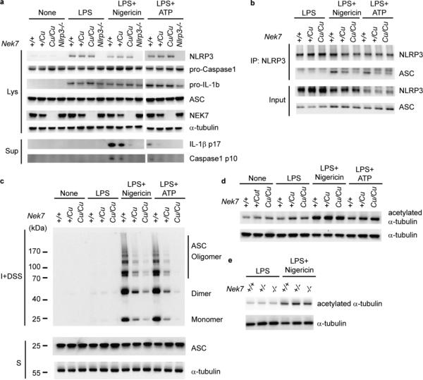
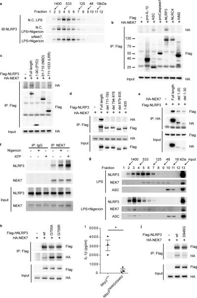
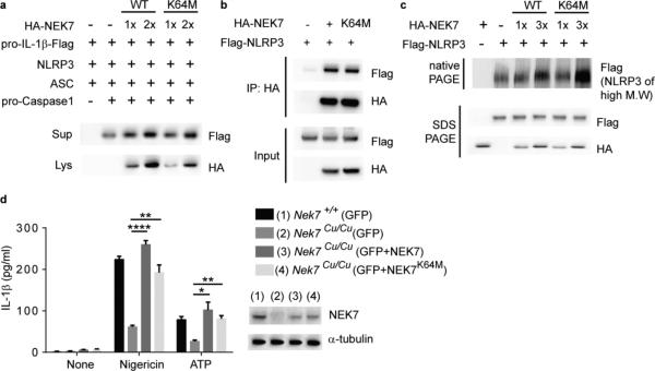
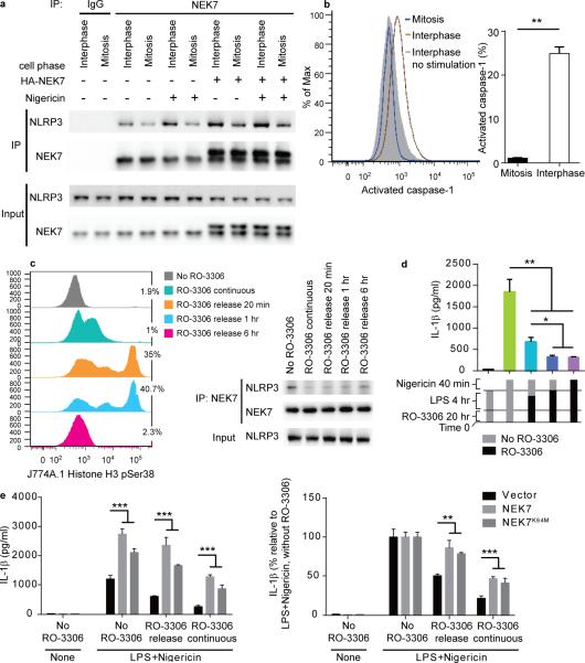
The NLRP3 inflammasome responds to microbes and danger signals by processing and activating proinflammatory cytokines, including interleukin 1β (IL-1β) and IL-18. We found here that activation of the NLRP3 inflammasome was restricted to interphase of the cell cycle by NEK7, a serine-threonine kinase previously linked to mitosis. Activation of the NLRP3 inflammasome required NEK7, which bound to the leucine-rich repeat domain of NLRP3 in a kinase-independent manner downstream of the induction of mitochondrial reactive oxygen species (ROS). This interaction was necessary for the formation of a complex containing NLRP3 and the adaptor ASC, oligomerization of ASC and activation of caspase-1. NEK7 promoted the NLRP3-dependent cellular inflammatory response to intraperitoneal challenge with monosodium urate and the development of experimental autoimmune encephalitis in mice. Our findings suggest that NEK7 serves as a cellular switch that enforces mutual exclusivity of the inflammasome response and cell division.


Figures

Figure 1
Figure 1
Impaired NLRP3 inflammasome activation in macrophages from Cuties mice. (a) ELISA analysis of IL-1β secretion by peritoneal macrophages from mice of the Cuties pedigree. Macrophages were primed with LPS and treated with nigericin. Data points represent individual mice; REF, Nek7+/+; HET, Nek7+/Cu; VAR, Nek7Cu/Cu. (b) Manhattan plot showing linkage of a mutation in Nek7 with the Cuties phenotype using a semidominant transmission model (P = 1.153 × 10⁻⁵). The −log10 P values are plotted versus the chromosomal positions of 79 mutations identified in the G1 founder of the pedigree. Horizontal red and purple lines represent thresholds of P = 0.05 with or without Bonferroni correction, respectively. (c) Immunoblot showing NEK7 expression in Cuties peritoneal macrophages. ELISA analysis of (d) IL-1β and (e) IL-18 secreted by peritoneal macrophages primed with LPS and treated with the indicated inflammasome stimuli. (f) Peritoneal macrophages were primed with LPS and treated with the indicated inflammasome stimuli. Pyroptosis, as measured by lactate dehydrogenase (LDH) released into the culture medium, is calculated relative to the total LDH activity in lysates of unstimulated cells. In d-f, n = 5 Nek7+/+, 5 Nek7+/Cu, 4 Nek7Cu/Cu, 4 Nlrp3−/− mice. * P≤0.05; ** P≤0.01; *** P≤0.001; **** P≤0.0001 (unpaired, two-tailed Student's t test). Results in d-f are representative of two independent experiments.
Figure 2
Figure 2
NEK7 promotes an inflammatory response in vivo. (a,b) Flow cytometric analysis of the number of peritoneal exudate cells (PECs), neutrophils (Ly6G+ F4/80), and monocytes/macrophages (F4/80+) in the peritoneum of (a) Cuties mutant mice or (b) bone marrow chimeric mice 6 h after intraperitoneal injection of MSU. For bone marrow chimeras, donor genotypes are indicated; recipients were Nek7+/+. Data points represent individual mice; the mean is indicated. (c) ELISA analysis of IL-1β in the lavage fluid of chimeric mice 6 h after MSU injection (n = 4 mice per genotype or chimera type). For bone marrow chimeras, donor genotypes are indicated; recipients were Nek7+/+. (d) Mean clinical score of mice immunized with rhMOG on day 0. P values indicate significance of differences relative to WT. (e) Flow cytometric analysis of the number of the indicated immune cell types in the spinal cords of mice 21 days after immunization with rhMOG (n = 4 mice per genotype). Cell subsets were gated as: NK (NK1.1+CD3ε−), monocytes/microglia (CD11b+Gr1), neutrophils (CD11b+Gr1+), lymphocytes (CD45hiCD11b). * P≤0.05; ** P≤0.01; *** P≤0.001; **** P≤0.0001 (unpaired, two-tailed Student's t test). Results in a-c are representative of two independent experiments. Results in d and e are representative of one experiment.
Figure 3
Figure 3
ROS, Ca2+ influx, and cAMP in Cuties macrophages in response to NLRP3 inflammasome stimuli. (a) Mitochondrial ROS. Flow cytometric analysis of MitoSOX staining in wild-type peritoneal macrophages primed with LPS (Mock), or macrophages of the indicated genotypes primed with LPS and treated with nigericin. (b) Calcium influx. Peritoneal macrophages were stained with calcium indicator fluo-4/AM. Images of untreated cells were acquired (t=0), then ATP was added to the culture medium and cells were imaged at 15 sec intervals for 30 min. The fold increase in fluorescence intensity of all cells in a field relative to time 0 is shown. (c) Intracellular cAMP in peritoneal macrophages primed with LPS and treated with nigericin or ATP. ** P≤0.01 (unpaired, two-tailed Student's t test). For a-c, n = 3 mice per genotype. Results are representative of two independent experiments.
Figure 4
Figure 4
NEK7 is necessary for NLRP3-ASC complex formation and ASC oligomerization. (a-c) Peritoneal macrophages were primed with LPS and stimulated with nigericin or ATP. Genotypes are for Nek7 except for lanes marked Nlrp3−/−. (a) Immunoblots showing expression of the indicated proteins in cell lysates (Lys) and culture supernatants (Sup). Secreted mature IL-1β p17 and active caspase-1 (p10 subunit) were measured in supernatants. (b) NLRP3-ASC association analyzed by immunoprecipitation and immunoblot. (c) Cell lysates were solubilized with Triton X-100-containing buffer. Insoluble (I) fractions were cross-linked with disuccinimidyl suberate (DSS) to capture ASC oligomers (I+DSS). The soluble (S) and I+DSS fractions were analyzed by immunoblotting with ASC antibodies. (d,e) Immunoblots showing acetylated α-tubulin and total α-tubulin in peritoneal macrophage lysates from (d) Cuties mice or (e) Nek7−/− mice. Results are representative of two independent experiments.
Figure 5
Figure 5
NEK7 directly interacts with the LRR domain of NLRP3 to promote inflammasome assembly. (a) RAW264.7 cells were transfected with non-targeting control siRNA (N.C.) or Nek7 siRNA (siNek7) and then primed with LPS and stimulated with nigericin. Immunoblot analysis of cell lysates subjected to gel filtration chromatography. (b-e) HEK293T cells were transfected as indicated. The interaction between HA-NEK7 and Flag-NLRP3 proteins was analyzed by immunoprecipitation and immunoblot. (d) Full length NLRP3 or LRR domain mutants lacking the indicated residues were tested. (e) Full length NEK7 or mutants lacking the indicated residues were tested. (f,g) Peritoneal macrophages were primed with LPS and stimulated with nigericin or ATP. (f) Endogenous NEK7-NLRP3 association in wild-type macrophages analyzed by immunoprecipitation and immunoblot. (g) Immunoblot analysis of wild-type macrophage lysates subjected to gel filtration chromatography. (h) HEK293T cells were transfected as indicated. Lysates were analyzed as in b. (i) ELISA analysis of IL-1β secretion by wild type or homozygous Nlrp3D946G mutant peritoneal macrophages primed with LPS and stimulated with nigericin. Data points represent individual mice. * P≤0.05 (unpaired, two-tailed Student's t test). (j) HEK293T cells were transfected as indicated. Lysates were analyzed as in b. Results are representative of two independent experiments.
Figure 6
Figure 6
NEK7 kinase activity is nonessential for NLRP3 inflammasome activation. (a-c) HEK293T cells were transfected as indicated. (a) Immunoblots showing pro-IL-1β-Flag and HA-NEK7 expression in cell lysates (Lys) and culture supernatants (Sup). IL-1β maturation was assessed by p17-Flag immunoblot of the supernatant. (b) The interaction between NEK7 or NEK7K64M and NLRP3 was analyzed by immunoprecipitation and immunoblot. (c) Flag immunoblot of cell lysates subjected to native PAGE to detect NLRP3 in high molecular weight complexes, or SDS-PAGE to detect total NLRP3. (d) Wild type or Nek7Cu/Cu peritoneal macrophages were electroporated with mRNA encoding GFP, GFP + NEK7, or GFP + NEK7K64M. GFP-positive cells were sorted, primed with LPS, and stimulated with nigericin or ATP. Secreted IL-1β was measured by ELISA. n = 3 mice per genotype. (Inset) NEK7 expression was assessed by immunoblot. * P≤0.05; ** P≤0.01; **** P≤0.0001 (unpaired, two-tailed Student's t test). Results are representative of two independent experiments.
Figure 7
Figure 7
NLRP3 inflammasome activation is blocked during mitosis. (a) NEK7-NLRP3 association in LPS-primed J774A.1 mitotic or interphase cells, with or without HA-NEK7 overexpression, analyzed by immunoprecipitation and immunoblot. (b) J774A.1 cells were primed with LPS and stimulated with nigericin together with fluorescent FAM-FLICA-caspase-1 probe specific for activated caspase-1. Flow cytometric analysis of cells containing activated caspase-1 (left); their frequency among mitotic and interphase cells is graphed (right). (c-e) J774A.1 cells were arrested at the G2/M phase border by incubation with RO-3306, then released from arrest for different time periods before analysis. (c) Cells were treated with RO-3306 for 20 h, with LPS added during the last 4 h. Release was in fresh media without RO-3306 and LPS for the indicated times. Flow cytometric analysis of phosphorylated histone H3-positive mitotic cells (left); the percentage of mitotic cells is indicated. The interaction between endogenous NEK7 and NLRP3 was analyzed by immunoprecipitation and immunoblot (right). (d) ELISA analysis of IL-1β in the culture supernatants of J774A.1 cells primed with LPS and stimulated with nigericin. Release from RO-3306 arrest was 1 h before (teal) or concurrent with (blue) nigericin stimulation. (e) ELISA analysis of IL-1β in the culture supernatants of J774A.1 cells stably overexpressing NEK7 or NEK7K64M primed with LPS and stimulated with nigericin. RO-3306 release was at the time nigericin was added. * P≤0.05; ** P≤0.01; *** P≤0.001 (unpaired, two-tailed Student's t test). For b, d, and e, the means of triplicate samples are plotted. Results are representative of two independent experiments.

Comment in

  • The NEK-sus of the NLRP3 inflammasome.
    Van Hauwermeiren F, Lamkanfi M. Van Hauwermeiren F, et al. Nat Immunol. 2016 Mar;17(3):223-4. doi: 10.1038/ni.3391. Nat Immunol. 2016. PMID: 26882252 No abstract available.

References

    1. Gross O, Thomas CJ, Guarda G, Tschopp J. The inflammasome: an integrated view. Immunol. Rev. 2011;243:136–151. - PubMed
    1. Zhou R, Yazdi AS, Menu P, Tschopp J. A role for mitochondria in NLRP3 inflammasome activation. Nature. 2011;469:221–225. - PubMed
    1. Subramanian N, Natarajan K, Clatworthy MR, Wang Z, Germain RN. The adaptor MAVS promotes NLRP3 mitochondrial localization and inflammasome activation. Cell. 2013;153:348–361. - PMC - PubMed
    1. Misawa T, et al. Microtubule-driven spatial arrangement of mitochondria promotes activation of the NLRP3 inflammasome. Nat. Immunol. 2013;14:454–460. - PubMed
    1. Roig J, Mikhailov A, Belham C, Avruch J. Nercc1, a mammalian NIMA-family kinase, binds the Ran GTPase and regulates mitotic progression. Genes Dev. 2002;16:1640–1658. - PMC - PubMed

Publication types

MeSH terms