

Rituximab does not reset defective early B cell tolerance checkpoints
- PMID: 26642366
- PMCID: PMC4701568
- DOI: 10.1172/JCI83840
Rituximab does not reset defective early B cell tolerance checkpoints
Abstract
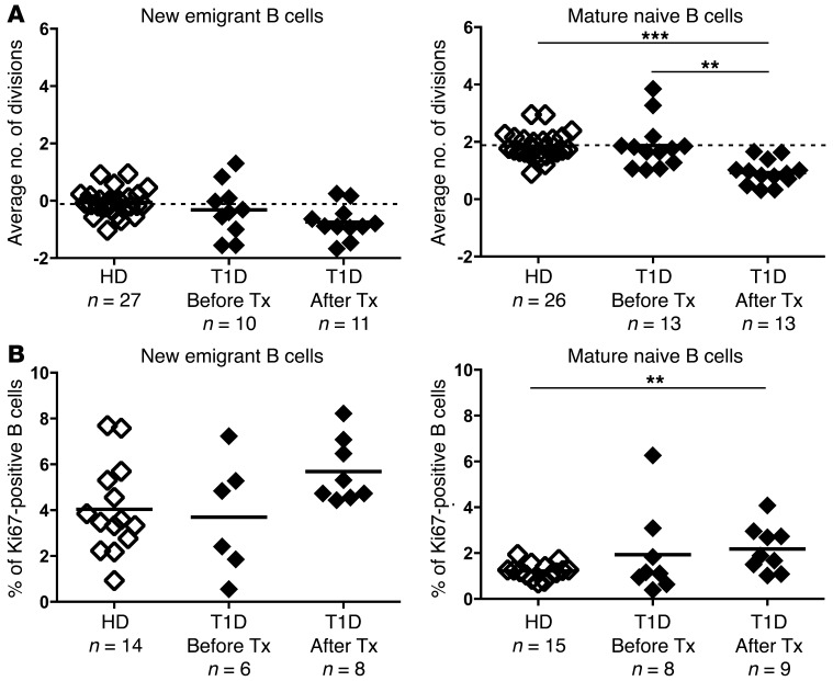
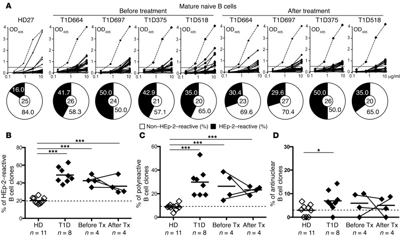
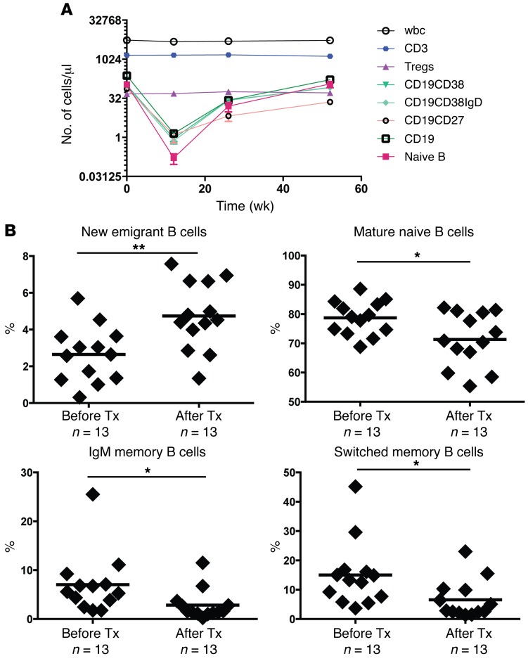
Type 1 diabetes (T1D) patients show abnormalities in early B cell tolerance checkpoints, resulting in the accumulation of large numbers of autoreactive B cells in their blood. Treatment with rituximab, an anti-CD20 mAb that depletes B cells, has been shown to preserve β cell function in T1D patients and improve other autoimmune diseases, including rheumatoid arthritis and multiple sclerosis. However, it remains largely unknown how anti-B cell therapy thwarts autoimmunity in these pathologies. Here, we analyzed the reactivity of Abs expressed by single, mature naive B cells from 4 patients with T1D before and 52 weeks after treatment to determine whether rituximab resets early B cell tolerance checkpoints. We found that anti-B cell therapy did not alter the frequencies of autoreactive and polyreactive B cells, which remained elevated in the blood of all patients after rituximab treatment. Moreover, the limited proliferative history of autoreactive B cells after treatment revealed that these clones were newly generated B cells and not self-reactive B cells that had escaped depletion and repopulated the periphery through homeostatic expansion. We conclude that anti-B cell therapy may provide a temporary dampening of autoimmune processes through B cell depletion. However, repletion with autoreactive B cells may explain the relapse that occurs in many autoimmune patients after anti-B cell therapy.
Figures
References
-
- Serreze DV, Fleming SA, Chapman HD, Richard SD, Leiter EH, Tisch RM. B lymphocytes are critical antigen-presenting cells for the initiation of T cell-mediated autoimmune diabetes in nonobese diabetic mice. J Immunol. 1998;161(8):3912–3918. - PubMed
Publication types
MeSH terms
Substances
Grants and funding
- U01 DK085476/DK/NIDDK NIH HHS/United States
- AI095848/AI/NIAID NIH HHS/United States
- U01 DK061010/DK/NIDDK NIH HHS/United States
- U01 DK103153/DK/NIDDK NIH HHS/United States
- U01 DK061058/DK/NIDDK NIH HHS/United States
- R01 AI071087/AI/NIAID NIH HHS/United States
- AI071087/AI/NIAID NIH HHS/United States
- U01 DK106984/DK/NIDDK NIH HHS/United States
- U01 DK085499/DK/NIDDK NIH HHS/United States
- U01 DK103266/DK/NIDDK NIH HHS/United States
- P01 AI061093/AI/NIAID NIH HHS/United States
- U01 DK107014/DK/NIDDK NIH HHS/United States
- AI082713/AI/NIAID NIH HHS/United States
- U19 AI082713/AI/NIAID NIH HHS/United States
- R21 AI095848/AI/NIAID NIH HHS/United States
- AI061093/AI/NIAID NIH HHS/United States
- U01 DK085465/DK/NIDDK NIH HHS/United States
LinkOut - more resources
Full Text Sources
Other Literature Sources
Medical
