Review
. 2016 Oct;134 Pt A(Pt A):145-161.
doi: 10.1016/j.nlm.2015.11.008. Epub 2015 Nov 28.

Prefrontal-hippocampal pathways underlying inhibitory control over memory

Affiliations
Review

Prefrontal-hippocampal pathways underlying inhibitory control over memory

Michael C Anderson et al. Neurobiol Learn Mem. 2016 Oct.

Abstract

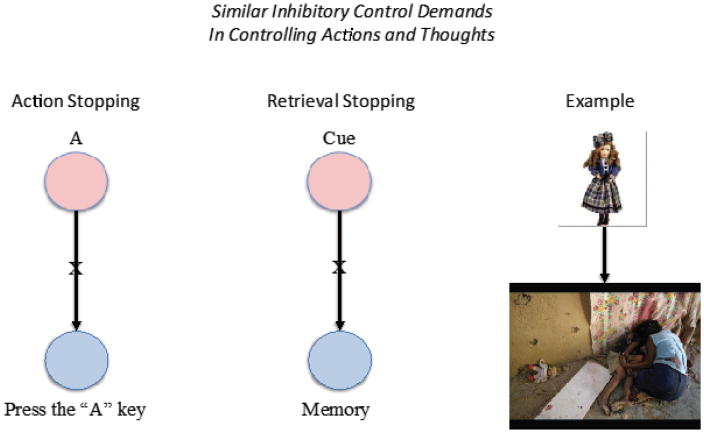
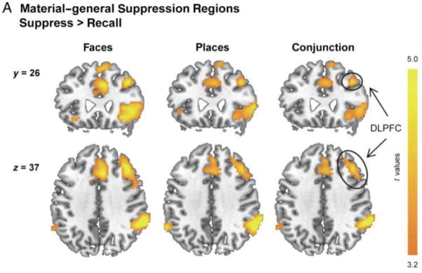
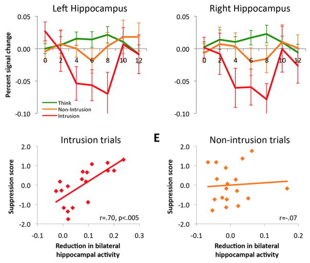
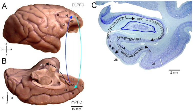
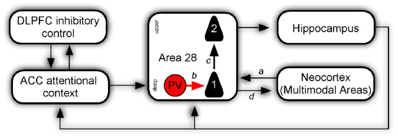
A key function of the prefrontal cortex is to support inhibitory control over behavior. It is widely believed that this function extends to stopping cognitive processes as well. Consistent with this, mounting evidence establishes the role of the right lateral prefrontal cortex in a clear case of cognitive control: retrieval suppression. Retrieval suppression refers to the ability to intentionally stop the retrieval process that arises when a reminder to a memory appears. Functional imaging data indicate that retrieval suppression involves top-down modulation of hippocampal activity by the dorsolateral prefrontal cortex, but the anatomical pathways supporting this inhibitory modulation remain unclear. Here we bridge this gap by integrating key findings about retrieval suppression observed through functional imaging with a detailed consideration of relevant anatomical pathways observed in non-human primates. Focusing selectively on the potential role of the anterior cingulate cortex, we develop two hypotheses about the pathways mediating interactions between lateral prefrontal cortex and the medial temporal lobes during suppression, and their cellular targets: the entorhinal gating hypothesis, and thalamo-hippocampal modulation via the nucleus reuniens. We hypothesize that whereas entorhinal gating is well situated to stop retrieval proactively, thalamo-hippocampal modulation may interrupt an ongoing act of retrieval reactively. Isolating the pathways that underlie retrieval suppression holds the potential to advance our understanding of a range of psychiatric disorders characterized by persistent intrusive thoughts. More broadly, an anatomical account of retrieval suppression would provide a key model system for understanding inhibitory control over cognition.

Keywords: Anterior cingulate; Forgetting; Hippocampus; Inhibitory control; Nucleus reuniens; Retrieval suppression.


Figures

Figure 1
Figure 1. Stopping actions and thoughts make similar inhibitory control demands
In a typical motor stopping task (left), a participant might receive a simple cue stimulus and be required to make an associated motor response as quickly as possible (e.g. seeing A and pressing the “A” key). On stop trials, people would be cued, mid-response to withhold the response (symbolized by the “X” on the association between the cue and response). Fulfilling this demands requires inhibitory control to suppress the motor action. Similarly, in a typical retrieval stopping situation, a stimulus appears in the world that is associated to a memory and that will lead us to be automatically reminded of the memory. If a person wishes to avoid being reminded, an inhibitory control process must be engaged to suppress retrieval of the associated memory (symbolized by the X on the association). In real life circumstances, retrieval suppression often arises after a trauma, when people seek to stop being reminded of unpleasant events (right side). For instance, after having witnessed an unpleasant scene (below), a later encounter with an object resembling something from the scene (top) has the power to elicit retrieval of the unpleasant event, triggering the need for control.
Figure 2
Figure 2. A typical set of suppression-related activations observed in a Think/No-Think study of retrieval suppression
Benoit et al. (2014) trained people on associations between words and faces, or between words and places. Displayed are brain areas that were significantly more activated when people suppressed (i.e., No-think trials) than when they retrieved items (Think trials), either when they were suppressing faces (left) or places (middle). The right side illustrates the conjunction analyses spanning these materials types, illustrating brain regions that generally are engaged during suppression, irrespective of the particular content. The strong right lateralization of activations is evident, as is the conspicuous involvement of the dorsolateral prefrontal cortex (DLPFC), ventrolateral prefrontal cortex (VLPFC), anterior cingulate cortex (ACC) and pre-supplementary motor area pre-SMA).
Figure 3
Figure 3. Illustration of hippocampal down-regulation during memory intrusions and its relationship to forgetting
Top Row; Activation in a priori structurally defined hippocampal regions of interest (ROIs) for Think trials and both types of No-Think trials: intrusions where the to-be-avoided memory entered awareness briefly and was purged, and non-intrusions where memory retrieval was successfully stopped. Note that whereas suppression reduces hippocampal activity in general, it does so more robustly for intrusions. Bottom Row, left; The magnitude of signal reduction in the hippocampus during intrusions (the average percentage signal change between 4 and 8 s after stimulus onset, displayed as a positive value) was correlated, across participants, with suppression-induced forgetting of No-Think items on the final test. Bottom row, right: This same measure of hippocampal activity during non-intrusions trials was not related to the amount of suppression-induced forgetting later observed. Error bars for all panels represent SEM.
Figure 4
Figure 4. Pathways linking the lateral and medial prefrontal cortices with the medial temporal lobe (MTL) memory system
A; Lateral surface of the rhesus monkey (Macaca mulatta) brain shows the location of Brodmann’s areas 9 (lateral), 46, frontopolar area 10 and areas 8 and 12. B; Medial surface of the brain shows the medial extent of areas 9 and 10, cingulate areas 24 and 32, and ventromedial areas 14 and 25. Lateral and medial prefrontal areas have robust bidirectional connections (cyan and blue arrows). The predominant projection to the medial temporal lobe (MTL) originates from the medial prefrontal areas in the anterior cingulate and terminates in the entorhinal (area 28) and perirhinal (area 35) cortices. C: Nissl stained coronal section through MTL of the monkey. Area 28 upper layers (II–III) originate the predominant cortical input to the hippocampus (HPC) where it is thought most mnemonic associations are made and contextually driven retrieval occurs. Hippocampal output first reaches the entorhinal deep layers (V–VI), which originate an ascending projection to the upper layers serving as a point by which signals can re-enter the hippocampal loop. Additionally, entorhinal deep layers originate the majority of cortico-cortical projections underlying the transfer of hippocampal signals to the perirhinal (areas 35, 36) and parahippocampal cortices (not shown).
Figure 5
Figure 5. Proposed ACC-Rhinal circuit underlying entorhinal gating
The diagram is simplified and shows only key pathways pertinent to inhibitory control. Lateral prefrontal cortex (DLPFC) shares bidirectional connections with medial prefrontal cortex (mPFC) in the anterior cingulate cortex (ACC), which is poised to exert inhibitory control on downstream structures in the medial temporal lobe (MTL). The ACC pathway to MTL forms synapses with excitatory neurons (not shown) as well as with parvalbumin (PV; red) putative inhibitory neurons in the deep layers of rhinal cortex (area 28). Through innervation of the powerful PV inhibitory neurons, the ACC may inhibit output of and input to the hippocampus. In the latter case, PV neurons would inhibit inputs to the ascending pathway (1) to the upper layers of area 28 (2), resulting in a loss of coordinated neural activity necessary to overcome rhinal inhibition, gating the hippocampus from incoming cortical information of sensory cues arriving from neocortical multimodal areas (a).
Figure 6
Figure 6. Proposed ACC-thalamic nucleus reuniens (RE) pathway underlying hippocampal modulation
A. The medial prefrontal cortex (mPFC) areas that receive robust projections from the hippocampus (HPC) (in the anterior cingulate cortex region) send a pathway to the RE that, in turn, originates one of the most prominent thalamic pathways to the medial temporal lobe (MTL), which terminate in CA1 as well as the subicular and rhinal cortices. B; Midsagittal drawing of the human brain denoting the position of three rostrocaudal levels through the RE which correspond to the three Nissl stained coronal sections (C, D, E).

References

    1. Andersen P, Holmqvist B, Voorhoeve PE. Entorhinal activation of dentate granule cells. Acta Physiologica Scandinavica. 1966;66:448–460. - PubMed
    1. Anderson MC, Green C. Suppressing unwanted memories by executive control. Nat. 2001;410(6826):131–134. - PubMed
    1. Anderson MC, Hanslmayr S. Neural mechanisms of motivated forgetting. Trends Cogn Sci. 2014;18:279–292. - PMC - PubMed
    1. Anderson MC, Huddleston E. Towards a Cognitive and Neurobiological Model of Motivated Forgetting. In: Belli RF, editor. True and false recovered memories: Toward a reconciliation of the debate. Vol. 58: Nebraska Symposium on Motivation. New York: Springer; 2011. - PubMed
    1. Anderson MC, Ochsner K, Kuhl B, Cooper J, Robertson E, Gabrieli SW, Glover G, Gabrieli JDE. Neural systems underlying the suppression of unwanted memories. Science. 2004;303:232–235. - PubMed