. 2016 Apr:145:130-139.
doi: 10.1016/j.exer.2015.11.017. Epub 2015 Nov 28.

Breakdown of interlocking domains may contribute to formation of membranous globules and lens opacity in ephrin-A5(-/-) mice

Affiliations

Breakdown of interlocking domains may contribute to formation of membranous globules and lens opacity in ephrin-A5(-/-) mice

Sondip Biswas et al. Exp Eye Res. 2016 Apr.

Abstract

Ephrin-A5, a ligand of the Eph family of receptor tyrosine kinases, plays a key role in lens fiber cell packing and cell-cell adhesion, with approximately 87% of ephrin-A5(-/-) mice develop nuclear cataracts. Here, we investigated the extensive formation of light-scattering globules associated with breakdown of interlocking protrusions during lens opacification in ephrin-A5(-/-) mice. Lenses from wild-type (WT) and ephrin-A5(-/-) mice between 2 and 21 weeks old were studied with light and electron microscopy, immunofluorescence labeling, freeze-fracture TEM and filipin cytochemistry for membrane cholesterol detection. Lens opacities with various densities were first observed in ephrin-A5(-/-) mice at around 60 days old. Dense cataracts in the mutant lenses were seen primarily in the nuclear region surrounded by transparent cortices from all eyes examined. We confirmed that a majority of nuclear cataracts were dislocated posteriorly and ruptured the thinner posterior lens capsule. SEM analysis indicated that numerous interlocking protrusions and wavy ridge-and-valley membrane surfaces in deep cortical and nuclear fibers did not cause lens opacity in both transparent ephrin-A5(-/-) and WT mice. In contrast, abundant isolated membranous globules of approximately 1000 nm in size were distributed randomly along the intact fiber cells during early stage of all ephrin-A5(-/-) cataracts examined. A further examination using both SEM and TEM revealed that isolated globules were generated from the disintegrated interlocking protrusions originally located along the corners of hexagonal fiber cells. Freeze-fracture TEM further revealed the association of square-array aquaporin junctions with both isolated globules and interlocking membrane domains. This study reports for the first time that disrupted interlocking protrusions are the source of numerous large membranous globules that contribute to light scattering and nuclear cataracts in the ephrin-A5(-/-) mice. Our results further suggest that dissociations of N-cadherin and adherens junctions in the associated interlocking domains may result in the formation of isolated globules and nuclear opacities in the ephrin-A5(-/-) mice.

Keywords: Adherens junction; Ephrin-A5 knockout mouse; Interlocking domain; Lens; Membranous globule; N-cadherin; Nuclear cataract.


Figures

Fig. 1
Fig. 1
Representative photographs of lens transparency and correlated morphologies in pairs of wild-type and ephrin-A5−/− lenses examined. (A) A pair of lenses from a wild-type mouse at age 12 weeks, which are transparent in both cortex (c) and nucleus (n). B–D show dense opacities in the lens nucleus in both lens pairs (B–C) or in only one lens (D) of ephrin-A5−/− mice at age 8–12 weeks. Note that these nuclear cataracts generally ruptured and protruded out of the posterior lens capsule based on our overview of the eyeballs. E–F show histological comparison between the intact wild-type lens and ruptured ephrin-A5−/− lens, in which the breakdown of lens nucleus into two fragmented portions (n–p1 and n–p2) is shown. Also noted is the nuclear fragment (n–p2) protruded considerably toward the posterior lens capsule. G–I show SEM comparison of intact fibers in wild-type and ruptured ephrin-A5−/− lenses at low magnifications. The similarity in structural regularity is observable between intact cortical fibers and nuclear fibers of a wild-type lens (G) in which several artificial damaged regions (arrows) during tissue processing are also shown. In contrast, significant irregularities in arrangements of deep cortical (dc) and nuclear fibers (n) in ephrin-A5−/− lenses (H and I) are evident. Scale bars: E–I = 1000 μm.
Fig. 2
Fig. 2
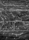
SEM comparison of interlocking protrusions in specific regions of cortical and nuclear fibers in wild-type and ephrin-A5−/− mice. The protrusions of normal cortical fiber cells change gradually from the smooth surface in the superficial cortex (A) and outer cortex (B) to ridge-and-valley surface in the deep cortex and nucleus (C, D), approximately from 100 μm to 650 μm deep from the equatorial lens surface of lenses in wild-type mice at age 8 weeks. The ridge-and-valley surface patterns, which represent wavy square-array junctions of fiber cells, are first seen in the inner cortex (C), approximately 400 μm deep from the lens surface, and are found extended toward the nuclear region of the lens (D). E–H shows changes in membrane surface and shape of protrusions in cortical and nuclear fibers in ephrin-A5−/− lenses. In the ephrin-A5−/− lenses, protrusions basically still retain their smooth membrane surface and regular shape in slightly irregular superficial fiber cells (e.g., 100 μm deep) as compared with the age-matched wild-type lenses (E). However, the dramatic changes of interlocking protrusions are seen in deeper cortex and nucleus which include irregular shape, size increase, and wavy ridge-and-valley surface patterns (F–H). These changes usually begin in outer cortex (~ 250 μm deep) and are extended toward the deep cortical and nuclear regions (G–H). Based on the dramatic changes in their shape (from elongated to round) and the size increase, it is conceivable that many of these protrusions might become the isolated protrusions or membrane-bound globules that are separated from their intact fiber cells. Scale bars: A–H = 1 μm.
Fig. 3
Fig. 3
SEM and light microscopy display a representative profile of interlocking protrusions and isolated membranous globules in damaged deep cortical fiber cells in ephrin-A5−/− mouse cataracts. A–D shows numerous round-shape protrusions or isolated globules of various sizes exhibiting either wavy ridge-and-valley surface pattern (arrows) or smooth surface (arrow heads). Light microscopy (E) shows distribution of numerous globules with different densities along the cell membranes, in the cytoplasm, or in the extracellular spaces. Scale bars: A–B = 10 μm; C–D = 1 μm; E = 30 μm.
Fig. 4
Fig. 4
Freeze-fracture TEM and filipin cytochemical analysis reveal the presence of intramembrane particles (proteins), square arrays and membrane cholesterol in isolated globules with either smooth or ridge-and-valley surface in ephrin-A5−/− lenses. Freeze-fracture TEM shows the presence of both e-face and p-face membranes on the same smooth globules (A), indicating that these globules are enclosed by double cell membranes. Note the 8–9 nm intramembrane particles are randomly distributed on the p-face of the membrane on the smooth globules (A). However, some isolated globules with undulating ridge-and-valley surfaces display different distribution patterns of intramembrane particles (B–C). They exhibit typical square-array configuration as those commonly seen in the deep cortical fibers (Biswas et al., 2014a; Biswas et al., 2014b). On these freeze-fracture surfaces, the p-face intramembrane particles of square arrays are generally distributed on the valley portion but not on the ridge portion (B and C). However, since the p-face square-array particles were often covered by the e-face membrane on the valley portion, these allowed only small rows of ~6.5 nm square-array particles along the sides of the valleys could sometime be observed (arrows). Filipin cytochemistry analysis reveals extensive distribution of membrane cholesterols (as represented by filipin-cholesterol-complexes, fcc) on both smooth and wavy isolated globules (D–F). However, the filipin-cholesterol-complexes are significantly decreased or absent in the patches of both flat (E) and wavy (F) square arrays (sa), due to the condensed localization of the square-array particles (Biswas et al., 2014b). Scale bars: A = 100 nm; B–F = 200 nm.
Fig. 5
Fig. 5
Thin-section TEM shows specific association of adherens junctions with interlocking protrusions in wild-type lenses. A low magnification overview of fiber cells reveals that adherens junctions (arrows) are regularly located at the corners of protrusions, or along the cell membranes near the apexes of fiber cells (A). B–C illustrates the specific localization of adherens junctions (arrows) associated with interlocking protrusions. High magnification reveals that intercellular adherens junctions are characterized as a spotty fascia-type (Lo, 1988; Lo et al., 2000) between lens fiber cells when they are clearly visualized under a favorable sectional orientation (D). Scale bars: A–B = 500 nm; C–D = 200 nm.
Fig. 6
Fig. 6
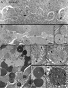
Thin-section TEM shows structural changes of interlocking protrusions and formation of membranous globules in ephrin-A5−/− lenses. (A) Although interlocking protrusions (p) still retain their normal-appearing configurations in superficial fiber cells, spotty adherens junctions were not readily visible at the corners of all protrusions shown. Arrows show several possible degraded spotty adherens junctions at the bottom of protrusions. In contrast, in the slightly deeper disorganized regions, many protrusions underwent significant shape changes into round or oval shapes with increasing sizes (B). Some were seen separated from the intact cell membrane (cm) to become isolated membrane-bound globules (C). High magnification reveals that representative isolated globules (g) contain light electron-density protein content, and are enclosed by double layers of wavy cell membranes (arrow heads) (D–E). The double cell membranes sometime form the unique aquaporin-0 thin junctions (arrows), approximately 12–14 nm in thickness (inset in D). A possible degraded adherens junction (open arrow) may also be seen associated with the membranous globule. In addition, in the much deeper cortex, the second type of isolated membranous globules (g), characterized by the dark electron densities, is often distributed randomly in the disorganized fiber cell regions (F–H). The isolated dark-density globules are most likely due to more accessible penetrations of osmium tetroxide and uranyl acetate stain into their contents, suggesting that they belong to the more damaging mature isolated type. High magnification also confirms that the isolated dark-density globules are bounded by double layers of cell membranes (arrow heads) which sometime form the unique aquaporin-0 thin junctions (arrows). Scale bars: A, C and G = 500 nm; B and F= 1000 nm; D, E and H = 200 nm. Inset in C = 20 nm.
Fig. 7
Fig. 7
Immunofluorescence labeling for the N-cadherin and β-catenin in wild type and ephrin-A5−/− mouse lenses at 12 weeks or 3 weeks old. The upper panel (A–F) shows significant decrease of N-cadherin immunoreactivity in fiber cell membrane from the superficial cortex to deep cortex in the ephrin-A5−/− lenses as compared with the wild type at 12 weeks old. The consecutive images were taken at the low magnification from the superficial to deep cortical fibers of cross sections along the equatorial plane. The approximate bottom location of each image (i.e., 120 μm, 260 μm and 400 μm) is indicated. The lower panel reveals at the high magnification that N-cadherin underwent early dislocation from the cortical fiber cells in the ephrin-A5−/− lenses at 3 weeks old (A–B). While the labeling for the N-cadherin (A) antibody is distributed evenly along the cortical fiber cell membranes in wild-type lens, the N-cadherin immunoreactivity is markedly released into cytoplasm of cortical fiber cells in ephrin-A5−/− lenses (B). However, the β-catenin labeling displays similar but lesser alterations (C–D). In addition, the labeling for EphA2 receptor antibody shows no change in cortical fiber cell membranes in both wild-type (C) and ephrin-A5−/− lenses (D). WGA is used here as a cell membrane marker. Scale bars: 20 μm for upper panel (A–F) and 5 μm for lower panel (A–D).
Fig. 8
Fig. 8
Immunofluorescence labeling on whole-mount samples for the N-cadherin in wild-type and ephrin-A5−/− mouse lenses at age 4 weeks. Whole-mount preparations of cortical fiber cells greatly facilitate visualization and labeling of interlocking protrusions. The labeling of the extracellular domains of N-cadherin antibody is clearly visualized along the protrusion membrane domains (arrows) in wild-type lens (A). This labeling pattern correlates well with the distribution of protrusions viewed on the SEM image (B). In contrast, the labeling of the extracellular domains of N-cadherin antibody is greatly defused into cytoplasm (arrows) of disorganized fiber cells in epherin-A5−/− mouse cataracts (C). SEM shows areas of enlarged extracellular spaces (arrows) in disorganized fiber cells of epherin-A5−/− lens (D). Scale bars: A and C = 5 μm; B and D = 1 μm.
Fig. 9
Fig. 9
A model of dissociation of the interlocking protrusions between lens fiber cells in the absence of ephrin-A5 function. A relatively normal configuration of elongated protrusions is regularly present between transparent superficial cortical fiber cells in ephrin-A5−/− lenses (A). The N-cadherin proteins (red bullets), and their associated adherens junctions, are richly associated with the protrusions. In the deeper cortical and nuclear regions, interlocking protrusions progressively undergo shape changes and dissociations from the cell membranes to form isolated membranous globules due to breakdown of N-cadherin-catenin complexes and associated adherens junctions in ephrin-A5−/− lenses (B–C). An accumulation of abundant isolated large globules at different maturation stages in the cells can cause light scatter and opacification in the deeper cortex and nucleus in ephrin-A5−/− lenses.

Similar articles

Cited by

References

    1. Atreya PL, Barnes J, Katar M, Alcala J, Maisel H. N-cadherin of the human lens. Curr Eye Res. 1989;8:947–956. - PubMed
    1. Bagchi M, Katar M, Lewis J, Maisel H. Associated proteins of lens adherens junction. J Cell Biochem. 2002;86:700–703. - PubMed
    1. Bhatnagar A, Ansari NH, Wang L, Khanna P, Wang C, Srivastava SK. Calcium-mediated disintegrative globulization of isolated ocular lens fibers mimics cataractogenesis. Exp Eye Res. 1995;61:303–310. - PubMed
    1. Biswas SK, Brako L, Gu S, Jiang JX, Lo WK. Regional changes of AQP0-dependent square array junction and gap junction associated with cortical cataract formation in the Emory mutant mouse. Exp Eye Res. 2014a;127:132–142. - PMC - PubMed
    1. Biswas SK, Brako L, Lo WK. Massive formation of square array junctions dramatically alters cell shape but does not cause lens opacity in the cav1-KO mice. Exp Eye Res. 2014b;125:9–19. - PMC - PubMed

Publication types