

Studying mechanisms of cAMP and cyclic nucleotide phosphodiesterase signaling in Leydig cell function with phosphoproteomics
- PMID: 26643407
- PMCID: PMC5679091
- DOI: 10.1016/j.cellsig.2015.11.014
Studying mechanisms of cAMP and cyclic nucleotide phosphodiesterase signaling in Leydig cell function with phosphoproteomics
Abstract
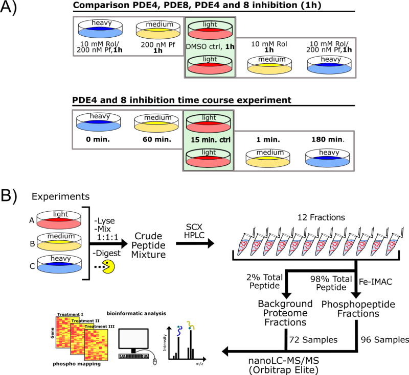
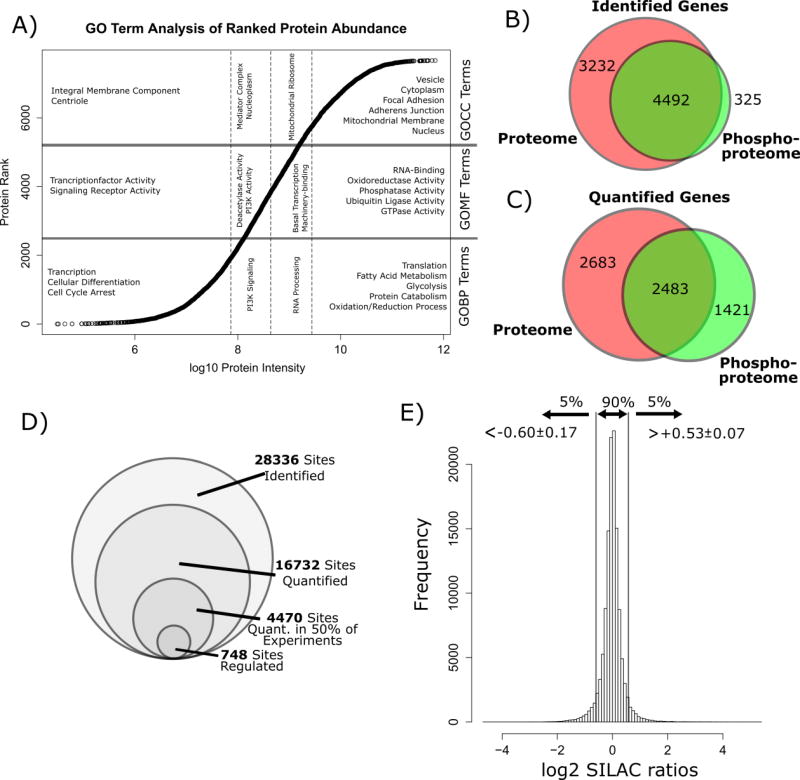
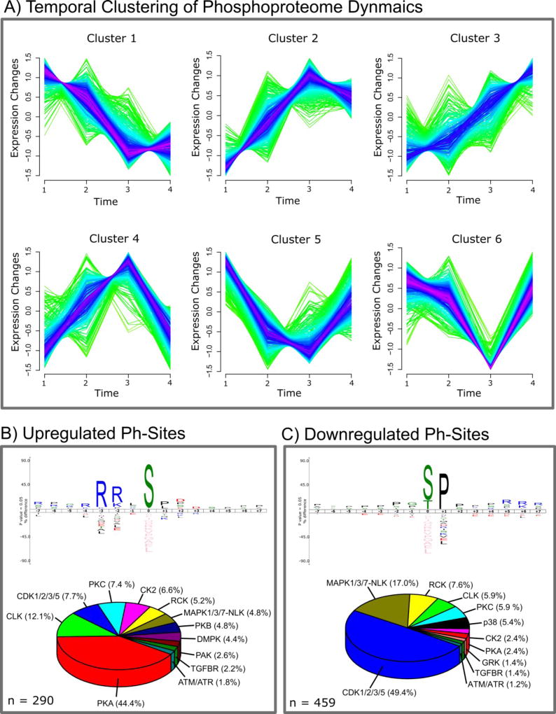
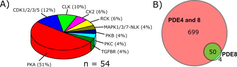
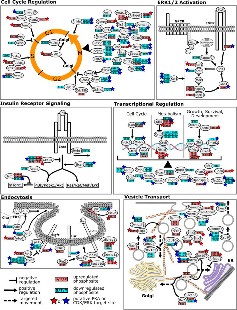
Many cellular processes are modulated by cyclic AMP and nucleotide phosphodiesterases (PDEs) regulate this second messenger by catalyzing its breakdown. The major unique function of testicular Leydig cells is to produce testosterone in response to luteinizing hormone (LH). Treatment of Leydig cells with PDE inhibitors increases cAMP levels and the activity of its downstream effector, cAMP-dependent protein kinase (PKA), leading to a series of kinase-dependent signaling and transcription events that ultimately increase testosterone release. We have recently shown that PDE4B and PDE4C as well as PDE8A and PDE8B are expressed in rodent Leydig cells and that combined inhibition of PDE4 and PDE8 leads to dramatically increased steroid biosynthesis. Here we investigated the effect of PDE4 and PDE8 inhibition on the molecular mechanisms of cAMP actions in a mouse MA10 Leydig cell line model with SILAC mass spectrometry-based phosphoproteomics. We treated MA10 cells either with PDE4 family specific inhibitor (Rolipram) and PDE8 family specific inhibitor (PF-04957325) alone or in combination and quantified the resulting phosphorylation changes at five different time points between 0 and 180min. We identified 28,336 phosphosites from 4837 proteins and observed significant regulation of 749 sites in response to PDE4 and PDE8 inhibitor treatment. Of these, 132 phosphosites were consensus PKA sites. Our data strongly suggest that PDE4 and PDE8 inhibitors synergistically regulate phosphorylation of proteins required for many different cellular processes, including cell cycle progression, lipid and glucose metabolism, transcription, endocytosis and vesicle transport. Our data suggests that cAMP, PDE4 and PDE8 coordinate steroidogenesis by acting on not one rate-limiting step but rather multiple pathways. Moreover, the pools of cAMP controlled by these PDEs also coordinate many other metabolic processes that may be regulated to assure timely and sufficient testosterone secretion in response to LH.
Keywords: Leydig cells; Phosphodiesterase; Phosphorylation; Proteomics; SILAC; Steroidogenesis.
Copyright © 2015 Elsevier Inc. All rights reserved.
Figures
Similar articles
-
cAMP-specific phosphodiesterases 8A and 8B, essential regulators of Leydig cell steroidogenesis.Mol Pharmacol. 2012 Apr;81(4):556-66. doi: 10.1124/mol.111.076125. Epub 2012 Jan 9. Mol Pharmacol. 2012. PMID: 22232524 Free PMC article.
-
Modulation of Leydig cell function by cyclic nucleotide phosphodiesterase 8A.Proc Natl Acad Sci U S A. 2006 Dec 26;103(52):19925-30. doi: 10.1073/pnas.0609483103. Epub 2006 Dec 15. Proc Natl Acad Sci U S A. 2006. PMID: 17172443 Free PMC article.
-
Src tyrosine kinase activity in rat thecal-interstitial cells and mouse TM3 Leydig cells is positively associated with cAMP-specific phosphodiesterase activity.Mol Cell Endocrinol. 1997 Jan 3;126(1):91-100. doi: 10.1016/s0303-7207(96)03975-5. Mol Cell Endocrinol. 1997. PMID: 9027367
-
The roles of cyclic nucleotide phosphodiesterases (PDEs) in steroidogenesis.Curr Opin Pharmacol. 2011 Dec;11(6):670-5. doi: 10.1016/j.coph.2011.09.003. Epub 2011 Sep 29. Curr Opin Pharmacol. 2011. PMID: 21962440 Free PMC article. Review.
-
cAMP-specific phosphodiesterase inhibitors: promising drugs for inflammatory and neurological diseases.Expert Opin Ther Pat. 2014 Dec;24(12):1311-21. doi: 10.1517/13543776.2014.968127. Epub 2014 Oct 4. Expert Opin Ther Pat. 2014. PMID: 25284693 Review.
Cited by
-
SCAP/SREBP pathway is required for the full steroidogenic response to cyclic AMP.Proc Natl Acad Sci U S A. 2016 Sep 20;113(38):E5685-93. doi: 10.1073/pnas.1611424113. Epub 2016 Sep 6. Proc Natl Acad Sci U S A. 2016. PMID: 27601673 Free PMC article.
-
An acquired scaffolding function of the DNAJ-PKAc fusion contributes to oncogenic signaling in fibrolamellar carcinoma.Elife. 2019 May 7;8:e44187. doi: 10.7554/eLife.44187. Elife. 2019. PMID: 31063128 Free PMC article.
-
Phosphatases modified by LH signaling in ovarian follicles: testing their role in regulating the NPR2 guanylyl cyclase†.Biol Reprod. 2024 Jan 13;110(1):102-115. doi: 10.1093/biolre/ioad130. Biol Reprod. 2024. PMID: 37774352 Free PMC article.
-
Roflumilast and aquaporin-2 regulation in rat renal inner medullary collecting duct.Physiol Rep. 2017 Jan;5(2):e13121. doi: 10.14814/phy2.13121. Physiol Rep. 2017. PMID: 28108651 Free PMC article.
-
H2 S catalysed by CBS regulates testosterone synthesis through affecting the sulfhydrylation of PDE.J Cell Mol Med. 2021 Apr;25(7):3460-3468. doi: 10.1111/jcmm.16428. Epub 2021 Mar 13. J Cell Mol Med. 2021. PMID: 33713531 Free PMC article.
References
-
- Beavo JA, Brunton LL. Cyclic nucleotide research -- still expanding after half a century. Nat Rev Mol Cell Biol. 2002;3:710–718. - PubMed
-
- Conti M, Beavo J. Biochemistry and physiology of cyclic nucleotide phosphodiesterases: essential components in cyclic nucleotide signaling. Annu. Rev. Biochem. 2007;76:481–511. - PubMed
-
- Hanoune J, Defer N. Regulation and role of adenylyl cyclase isoforms. Annu. Rev. Pharmacool. Toxicol. 2001;41:145–174. - PubMed
Publication types
MeSH terms
Substances
Grants and funding
LinkOut - more resources
Full Text Sources
Other Literature Sources
