. 2016 Jan 15;196(2):543-6.
doi: 10.4049/jimmunol.1502080. Epub 2015 Dec 7.

Cutting Edge: IL-1 Receptor Signaling is Critical for the Development of Autoimmune Uveitis

Affiliations

Cutting Edge: IL-1 Receptor Signaling is Critical for the Development of Autoimmune Uveitis

Chi-Keung Wan et al. J Immunol. .

Abstract

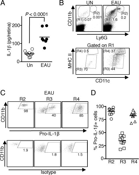
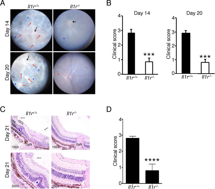
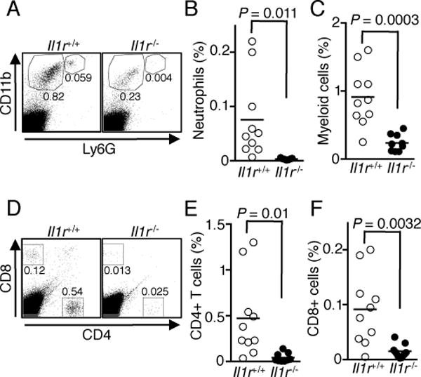
IL-1β is a proinflammatory cytokine important for local and systemic immunity. However, aberrant production of this cytokine is implicated in pathogenic mechanisms of a number of inflammatory diseases, including Behçet's disease and age-related macular degeneration. In this study, we report the increased secretion of IL-1β in the retina by neutrophils, macrophages, and dendritic cells during ocular inflammation and show that loss of IL-1R signaling confers protection from experimental autoimmune uveitis. Moreover, the amelioration of experimental autoimmune uveitis in Il1r-deficient mice was associated with reduced infiltration of inflammatory cells into the retina and decreased numbers of uveitogenic Th17 cells that mediate uveitis. These findings indicate the possible utility of IL-1R-blocking agents for the treatment of ocular inflammatory diseases.


Figures

FIGURE 1
FIGURE 1
IL-1β is expressed in the retina during EAU. Retinae of WT mice without immunization (Un) and with EAU were isolated on day 21 post-immunization, digested with collagenase and analyzed by ELISA (A) or flow cytometry (B–D). (A) Analysis of IL-1β secretion by ELISA. Data are from 2 independent experiments with a total of 7–10 mice. (B) Macrophages/DCs (CD11b+ Ly6G, R1) and neutrophils (CD11b+ Ly6G+, R2) that had infiltrated into the retina were first identified by CD11b and Ly6G staining. Macrophages and DCs were defined as CD11c+ MHC IIlow macrophages (R3) and CD11chi MHC IIhi DCs (R4) (C and D) Expression of intracellular pro-IL-1β in the immune cells identified in Fig. 1B after EAU induction. Shown are representative flow cytometric plots (C) and as well as data from 10 individual mice (D).
FIGURE 2
FIGURE 2
Il1r-deficient mice develop less severe EAU. EAU was induced in WT and Il1r−/− mice. (A) Fundus images of the eyes at days 14 and 20 after EAU induction. Compared to the Il1r−/− mice, the retinae of WT mice reveals obvious inflammation with blurred optic disc margins and enlarged juxtapapillary area (black arrows), retinal vasculitis with moderate or severe cuffing (red arrows), and yellow-whitish retinal and choroidal infiltrates (blue arrows). (B) Clinical score and assessment of disease severity were based on changes at the optic nerve disc or retinal vessels and retinal and choroidal infiltrates as described in the text. ***, P < 0.001. (C) Histological analysis of the retina at day 21 after EAU induction shows increased numbers of inflammatory cells in the vitreous (black arrows), retinal folds (blue asterisk) in retina of WT compared to Il1r−/− mice. (D) Significant reduction of histological score in Il1r−/− mice compared to WT mice. Data are representative of 2 independent experiments (total of 10 mice/group). ****, P < 0.0001. Sections were stained with H&E staining. V, vitreous, black arrow, infiltrated for inflammatory cells, Blue asterisk, retinal folds; OpN, optic nerve; GCL, ganglion cell layer; INL, inner nuclear layer; ONL outer nuclear layer; RPE/CH retinal pigment epithelial cell layer; and choroid.
FIGURE 3
FIGURE 3
Il1r−/− mice have fewer immune cells infiltrating into the retina during EAU. EAU was induced in WT and Il1r−/− mice, retinae were isolated at day 21 post-immunization, digested with collagenase and analyzed by flow cytometry. (A) Shown are representative blots of CD11b versus Ly6G staining. (B and C) Percentages of neutrophils (B) and macrophages/DCs (C) from individual mice. (D) Shown are representative blots of CD4 and CD8 staining. (E and F) Percentages of CD4+ (E) and CD8+ (F) T cells. Data shown are from 2 independent experiments with a total of 10 mice/group.
FIGURE 4
FIGURE 4
IL-1R-dependent signaling is required for Th17 cell differentiation. (A–C) EAU was induced in WT and Il1r−/− mice, retinae were isolated on day 21 post-immunization and analyzed by the intracellular cytokine staining assay. Shown are representative flow cytometric plots (A) and percentages of CD4+ T cells expressing IL-17 (B) and IFN-γ (C) from individual mice. (D and E) Cervical and draining lymph node cells were re-stimulated with IRBP1–20 peptide for 3 d. Amounts of IL-17 (D), IFN-γ (E), and IL-1β (F) in the culture were determined by ELISA. Data shown are from 2 independent experiments with a total of 10 mice/group.

References

    1. Amadi-Obi A, Yu CR, Liu X, Mahdi RM, Clarke GL, Nussenblatt RB, Gery I, Lee YS, Egwuagu CE. TH17 cells contribute to uveitis and scleritis and are expanded by IL-2 and inhibited by IL-27/STAT1. Nature medicine. 2007;13:711–718. - PubMed
    1. Wang RX, Yu CR, Dambuza IM, Mahdi RM, Dolinska MB, Sergeev YV, Wingfield PT, Kim SH, Egwuagu CE. Interleukin-35 induces regulatory B cells that suppress autoimmune disease. Nature medicine. 2014;20:633–641. - PMC - PubMed
    1. van de Veerdonk FL, Netea MG, Dinarello CA, Joosten LA. Inflammasome activation and IL-1beta and IL-18 processing during infection. Trends in immunology. 2011;32:110–116. - PubMed
    1. Gabay C, Lamacchia C, Palmer G. IL-1 pathways in inflammation and human diseases. Nature reviews Rheumatology. 2010;6:232–241. - PubMed
    1. Chung Y, Chang SH, Martinez GJ, Yang XO, Nurieva R, Kang HS, Ma L, Watowich SS, Jetten AM, Tian Q, Dong C. Critical regulation of early Th17 cell differentiation by interleukin-1 signaling. Immunity. 2009;30:576–587. - PMC - PubMed

Publication types