. 2015 Oct-Dec;4(4):312-8.
doi: 10.4103/2303-9027.170423.

Morphological differentiation and follow-up of pancreatic cystic neoplasms using endoscopic ultrasound

Affiliations

Morphological differentiation and follow-up of pancreatic cystic neoplasms using endoscopic ultrasound

Susumu Hijioka et al. Endosc Ultrasound. 2015 Oct-Dec.

Abstract

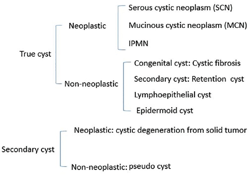
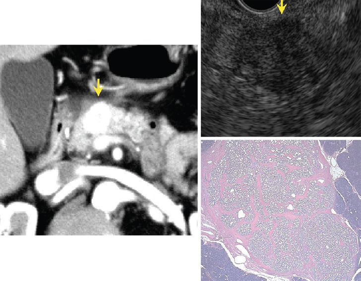
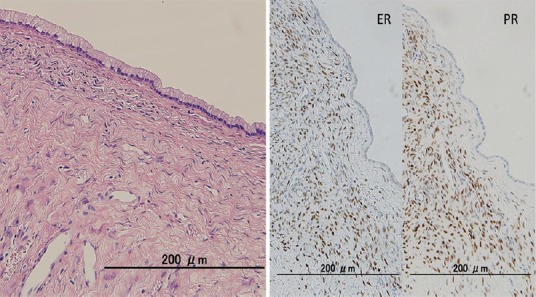
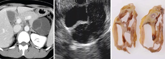
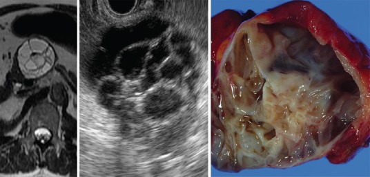
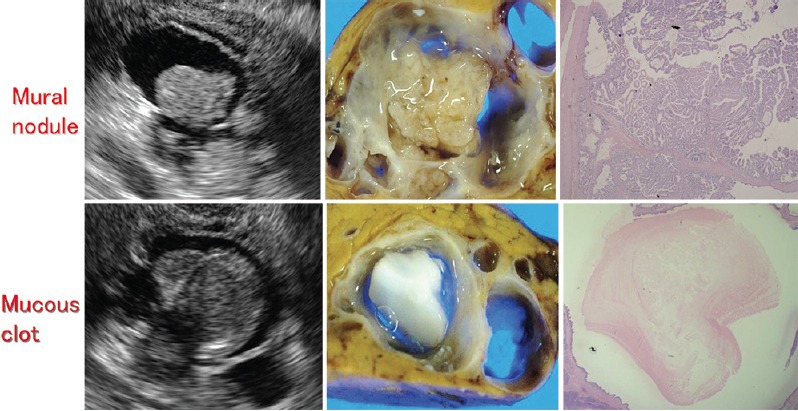
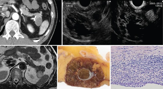
Endoscopic ultrasound (EUS) is a key modality for the evaluation of suspected pancreatic cystic neoplasms (PCNs), as the entire pancreatic gland can be demonstrated with high spatial resolution from the stomach and duodenum. Detailed information can be acquired about the internal contents of the cyst(s) [septum, capsule, mural nodules (MNs)], its relation with the main pancreatic duct (MPD), and any parenchymal changes in the underlying gland. PCNs comprise true cysts and pseudocysts. True cysts can be neoplastic or nonneoplastic. Here, we describe serous cystic neoplasm (SCN), mucinous cystic neoplasm (MCN), and intraductal papillary mucinous neoplasm (IPMN) as prototype neoplastic cysts, along with nonneoplastic lymphoepithelial cysts (LECs).


Figures

Figure 1
Figure 1
Classification of pancreatic cystic neoplasms
Figure 2
Figure 2
Classification of serous cystic neoplasms (SCNs)
Figure 3
Figure 3
Microcystic type serous cystic neoplasms (SCNs). EUS image (arrow) shows honeycomb-like appearance caused microcyst accumulation
Figure 4
Figure 4
Macrocystic type and microcystic type serous cystic neoplasms (SCNs)
Figure 5
Figure 5
Solid type serous cystic neoplasm (SCN). Enhancement is intense on CT. Honeycomb-like appearance is undetectable by EUS but pathological microscopic findings show small microcystic appearance
Figure 6
Figure 6
Ovarian type stroma (OTS) immediately below the epithelium detected by microscopy is estrogen-receptor and progesterone-receptor positive
Figure 7
Figure 7
Image of MCN adenoma shows round macrocystic lesion with thickened wall and septum
Figure 8
Figure 8
Image of MCN adenoma shows cysts within cysts
Figure 9
Figure 9
Mural cyst of MCN adenoma. Apparent mural nodule in CT image appearing as a mural cyst in EUS image is MCN
Figure 10
Figure 10
Branch duct (BD) type IPMN (noninvasive carcinoma). Hyperechoic mass (arrow) in dilated branch is evident on EUS and IDUS images. Contrast agents show blood flow signals in mass indicating mural nodules
Figure 11
Figure 11
Upper and lower panels show mural nodule (adenoma) and mucous clot, respectively
Figure 12
Figure 12
MD type IPMN (noninvasive carcinoma). Mural nodule is detectable inside MPD
Figure 13
Figure 13
Lymphoepithelial cysts

References

    1. Sakorafas GH, Smyrniotis V, Sarr MG. Pancreatic cystic neoplasms. 2015
    1. Hocke M, Cui XW, Domagk D, et al. Pancreatic cystic lesions: The value of contrast-enhanced endoscopic ultrasound to influence the clinical pathway. Endosc Ultrasound. 2014;3:123–30. - PMC - PubMed
    1. Lennon AM, Wolfgang C. Cystic neoplasms of the pancreas. J Gastrointest Surg. 2013;17:645–53. - PubMed
    1. Barresi L, Tarantino I, Granata A, et al. Pancreatic cystic lesions: How endoscopic ultrasound morphology and endoscopic ultrasound fine needle aspiration help unlock the diagnostic puzzle. World J Gastrointest Endosc. 2012;4:247–59. - PMC - PubMed
    1. Cho CS, Russ AJ, Loeffler AG, et al. Preoperative classification of pancreatic cystic neoplasms: The clinical significance of diagnostic inaccuracy. Ann Surg Oncol. 2013;20:3112–9. - PMC - PubMed