

Cerebral reactivity in migraine patients measured with functional near-infrared spectroscopy
- PMID: 26644117
- PMCID: PMC4672549
- DOI: 10.1186/s40001-015-0190-9
Cerebral reactivity in migraine patients measured with functional near-infrared spectroscopy
Abstract
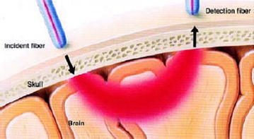
Background: There are two major theories describing the pathophysiology of migraines. Vascular theory explains that migraines resulted from vasodilation of meningeal vessels irritating the trigeminal nerves and causing pain. More recently, a neural theory of migraine has been proposed, which suggests that cortical hyperexcitability leads to cortical spreading depression (CSD) causing migraine-like symptoms. Chronic migraine requires prophylactic therapy. When oral agents fail, there are several intravenous agents that can be used. Understanding underlying causes of migraine pain would help to improve efficacy of migraine medications by changing their mechanism of action. Yet to date no study has been made to investigate the link between vascular changes in response to medications for migraine versus pain improvements. Functional near-infrared spectroscopy (NIRS) has been used as an inexpensive, rapid, non-invasive and safe technique to monitor cerebrovascular dynamics.
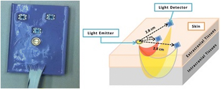
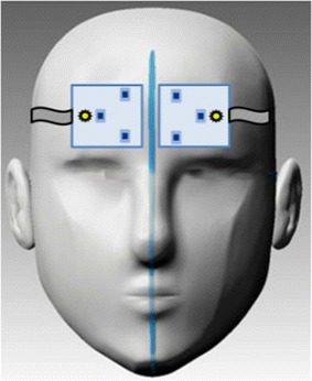
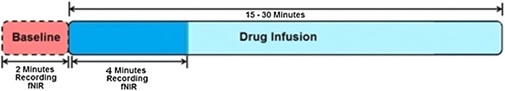
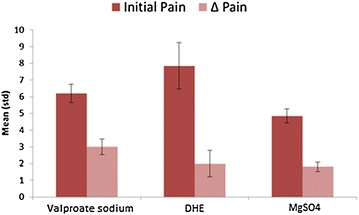
Method: In this study, a multi-distance near-infrared spectroscopy device has been used to investigate the cortical vascular reactivity of migraine patients in response to drug infusions and its possible correlation with changes in pain experienced. We used the NIRS on 41 chronic migraine patients receiving three medications: magnesium sulfate, valproate sodium, and dihydroergotamine (DHE). Patients rated their pain on a 1-10 numerical scale before and after the infusion.
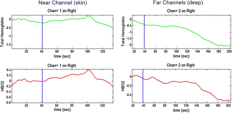
Results: No significant differences were observed between the medication effects on vascular activity from near channels measuring skin vascularity. However, far channels--indicating cortical vascular activity--showed significant differences in both oxyhemoglobin and total hemoglobin between medications. DHE is a vasoconstrictor and decreased cortical blood volume in our experiment. Magnesium sulfate has a short-lived vasodilatory effect and increased cortical blood volume in our experiment. Valproate sodium had no significant effect on blood volume. Nonetheless, all three reduced patients' pain based on self-report and no significant link was observed between changes in cortical vascular reactivity and improvement in migraine pain as predicted by the vascular theory of migraine.
Conclusion: NIRS showed the potential to be a useful tool in the clinical setting for monitoring the vascular reactivity of individual patients to various migraine and headache medications.
Figures
References
-
- Goadsby PJ. The vascular theory of migraine—a great story wrecked by the facts. Brain J Neurol. 2009;132(Pt 1):6–7. - PubMed
-
- Sayita Y (2012) Classification of migraineurs using functional near infrared spectroscopy data. The middle east technical university
MeSH terms
Substances
LinkOut - more resources
Full Text Sources
Other Literature Sources
Medical
