. 2016 Sep 25;33(5 Pt A):551-64.
doi: 10.1016/j.nbt.2015.11.007. Epub 2015 Nov 28.

Developing high-quality mouse monoclonal antibodies for neuroscience research - approaches, perspectives and opportunities

Affiliations

Developing high-quality mouse monoclonal antibodies for neuroscience research - approaches, perspectives and opportunities

Belvin Gong et al. N Biotechnol. .

Abstract

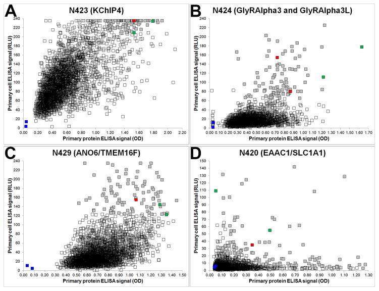
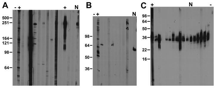
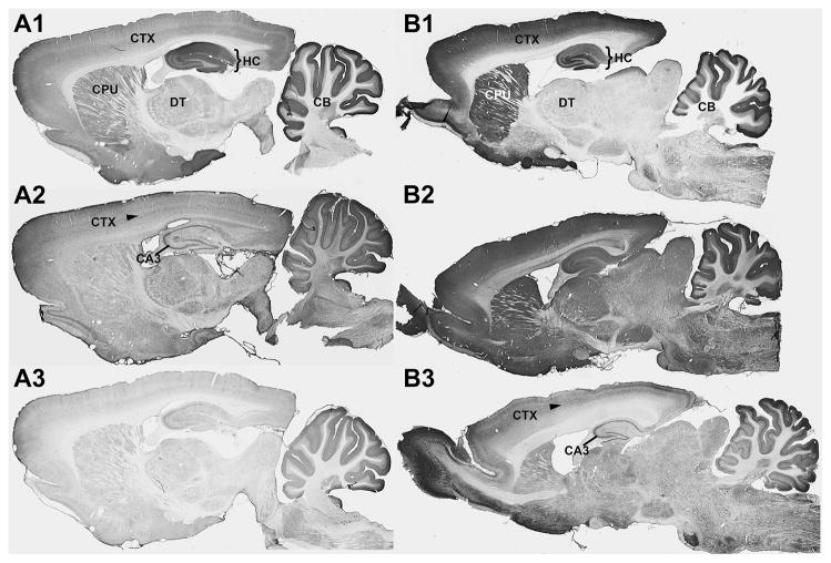
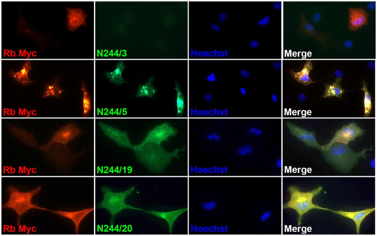
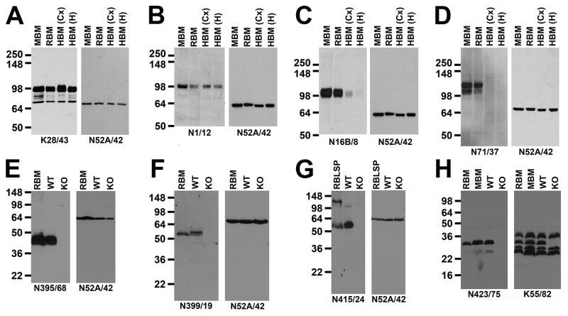
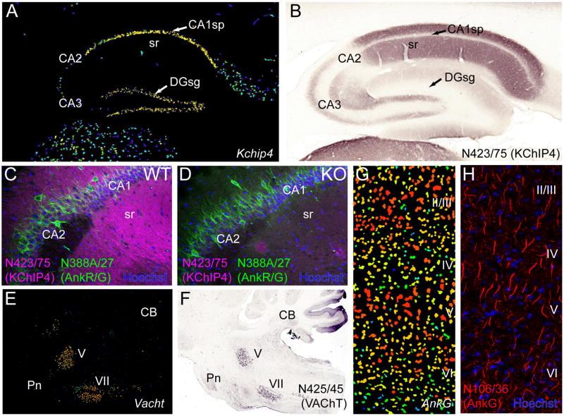
High-quality antibodies (Abs) are critical to neuroscience research, as they remain the primary affinity proteomics reagent used to label and capture endogenously expressed protein targets in the nervous system. As in other fields, neuroscientists are frequently confronted with inaccurate and irreproducible Ab-based results and/or reporting. The UC Davis/NIH NeuroMab Facility was created with the mission of addressing the unmet need for high-quality Abs in neuroscience research by applying a unique approach to generate and validate mouse monoclonal antibodies (mAbs) optimized for use against mammalian brain (i.e., NeuroMabs). Here we describe our methodology of multi-step mAb screening focused on identifying mAbs exhibiting efficacy and specificity in labeling mammalian brain samples. We provide examples from NeuroMab screens, and from the subsequent specialized validation of those selected as NeuroMabs. We highlight the particular challenges and considerations of determining specificity for brain immunolabeling. We also describe why our emphasis on extensive validation of large numbers of candidates by immunoblotting and immunohistochemistry against brain samples is essential for identifying those that exhibit efficacy and specificity in those applications to become NeuroMabs. We describe the special attention given to candidates with less common non-IgG1 IgG subclasses that can facilitate simultaneous multiplex labeling with subclass-specific secondary antibodies. We detail our recent use of recombinant cloning of NeuroMabs as a method to archive all NeuroMabs, to unambiguously define NeuroMabs at the DNA sequence level, and to re-engineer IgG1 NeuroMabs to less common IgG subclasses to facilitate their use in multiplex labeling. Finally, we provide suggestions to facilitate Ab development and use, as to design, execution and interpretation of Ab-based neuroscience experiments. Reproducibility in neuroscience research will improve with enhanced Ab validation, unambiguous identification of Abs used in published experiments, and end user proficiency in Ab-based assays.


Figures

Figure 1
Figure 1. Representative ELISA primary screen data
Scatter plots show the relative distribution of ELISA data from NeuroMab projects N423 (A), N424 (B), N429 (C) and N420 (D), where protein and cell ELISA data are plotted on the x- and y-axis, respectively. Each project began with 2,944 hybridoma-containing wells and each graph shows 2,948 data points (squares) that together comprise positive (green) and negative (blue) control wells (each the average of 32 replicates), unselected wells (unfilled squares) and selected wells (grey squares), with red squares denoting the wells with candidates that ultimately became NeuroMabs.
Figure 2
Figure 2. Representative immunoblot secondary screen data
Images show immunoblot data from three different NeuroMab projects. (A) Anti-pan Nav1 channel N419 NeuroMab project. (B) Anti-GABA(A) receptor π subunit N431 NeuroMab project. (C) Anti-KChIP4 N423 NeuroMab project. Numbers to the left of each panel show the mobility of molecular weight standards in kDa. Adult rat brain protein samples analyzed were a P2 crude membrane fraction or RBM (A and C) or a P1 low-speed pellet or RBLSP (B). Expected relative electrophoretic mobilities for target bands were 250 kDa (A), 50 kDa (B), and 30 kDa (C). The subset of strips shown depicts multiple candidates. (A) N419/28 to N419/40. (B) N431/55 to N431/64. (C) N423/28 to N423/37. Each set includes respective positive and negative controls (plus and minus symbols, respectively). The “N” denotes candidates that ultimately became a NeuroMab. (A) N419/40. (B) N431/64. (C) N423/75.
Figure 3
Figure 3. Representative images from IHC secondary screens
Photomicrographs show DAB/NAS immunolabeling of sagittal sections of adult rat brain from NeuroMab projects N355 (A1, A2 and A3) and N399 (B1, B2 and B3). There were 96 candidates screened for each project and each column shown here depicts images collected from one candidate judged to meet the criteria set out for specific labeling (N355/1 in A1 and N399/19 in B1) and two other candidates from these projects that failed to meet these criteria (N355/2 in A2, N355/3 in A3, N399/17 in B2 and N399/18 in B3). The bracketed region in A1 and B1 is the hippocampus, and the arrow in A2 and B3 identifies neocortex layer 5. CA3, hippocampal cornu ammonis area 3, CB, cerebellum, CPU, caudoputamen, CTX, neocortex DT, dorsal thalamus, HC, hippocampus.
Figure 4
Figure 4. Representative images from IF-ICC secondary screens
Photomicrographs show IF immunolabeling of COS-1 cells transiently transfected with a Myc-tagged rat SynCAM4 mammalian expression construct using a rabbit anti-Myc pAb (red), candidate mouse mAbs (green) and Hoechst nuclear stain (blue). Each row depicts a set of images collected from a different candidates, one presented as typical of a negative candidate (N244/3), and three represented as positive candidates (N244/5, N244/19 and N244/20), of which N244/5 ultimately became a NeuroMab.
Figure 5
Figure 5. Tertiary screening by IB against samples from human brain, and from KO mouse brain
Panels (A–D) depict results from NeuroMab TCS screening against mouse, rat and human brain P2 crude membrane fraction samples (MBM: adult mouse brain; RBM: adult rat brain; HBM (adult human brain, from Cx: neocortex; Hi: hippocampus). (A) Anti-PSD-95 NeuroMab K28/43. (B) Anti-KCC2 NeuroMab N1/12. (C) Anti-Kv3.1b NeuroMab N16B/8. (D) Anti-HCN2 NeuroMab N71/37. Panels (E–H) depict results from NeuroMab TCS screening against brain samples from WT mice, and from the KO mouse corresponding to the NeuroMab target. Samples in panels (E, F) and (H) are P2 crude membrane fractions from adult rat brain (RBM), or adult mouse brains from WT and KO mice. Panel (G) has P1 low-speed pellet fractions from adult rat brain (RBLSP), or from adult WT and KO mouse brains. (E) Anti-Navβ2 subunit NeuroMab N395/68. (F) Anti-GABA(A) receptor α2 subunit NeuroMab N399/19. (G) Anti-GABA(A) receptor α5 subunit NeuroMab N415/24. (H) Anti-KChIP4 Kv channel subunit NeuroMab N423/75. In all cases duplicate immunoblots were probed the anti-Mortalin/GRP75 NeuroMab N52A/42 as a control to show comparable loading of all samples, with the exception of (H) in which the anti-pan-KChIP NeuroMab K55/82 was used to show the selective elimination of the KChIP4 band within the brain KChIP population in the KChIP4 KO sample. Numbers to the left of each panel show the mobility of molecular weight standards in kDa.
Figure 6
Figure 6. Tertiary screening by IHC
Panels (A, B) show that discordance between regional localization of transcript and protein in mammalian brain can confound simple Ab evaluation. (A) Pseudocolor image of a sagittal section from mouse brain processed for in situ hybridization to mRNA for Kchip4 obtained from the ABA (image 70546039_100) showing prominent transcript expression in the principal cell layers of the hippocampus, where cell bodies reside. (B) A sagittal section from rat brain immunolabeled for KChIP4 using NeuroMab N423/75 TCS by nickel enhanced diaminobenzidine (NiDAB) histochemistry showing a lack of labeling in the principal cellular layers of the hippocampus, but prominent labeling in the molecular layers, particularly stratum radiatum (sr) which is rich in dendrites. (C, D) Antibody specificity confirmed by KO validation. (C) IF labeling for KChIP4 using NeuroMab N423/75 TCS (magenta) in a sagittal section from a WT mouse brain shows the same prominent immunolabeling pattern in the dendrite rich molecular layers of hippocampus observed in rat (B). (D) Immunolabeling for KChIP4 is absent in the KChIP4 KO brain. In contrast, immunolabeling with the anti-ankyrin-R/G NeuroMab N388A/27 TCS (green) does not differ between WT and KO. Dye staining with Hoechst 33342 (blue) labels cellular nuclei. (E, F) Cellular specificity of transcript expression can guide Ab evaluation. (E) Pseudocolor image of in situ hybridization to VAChT mRNA in a sagittal mouse section from the ABA (image 79762433_86) showing specific transcript expression in the cranial trigeminal motor nucleus (V) and facial nucleus (VII) of the brainstem. (F) Immunolabeling by NiDAB histochemistry using NeuroMab N425/45 TCS in a sagittal section through the brainstem of a rat shows prominent signal for VAChT in the same cell nuclei (V, VII) as mRNA. (G,H) Subcellular target localization can guide NeuroMab evaluation. (G) Pseudocolor image of in situ hybridization of Ankyrin-G mRNA from the ABA (image 68196979_98) showing intense cellular expression throughout all cortical laminae (II–VI). (H) IF labeling for Ankyrin-G (magenta) with NeuroMab N106/36 TCS and staining against cellular nuclei with the dye Hoechst 33342 (blue). In contrast to mRNA, which is localized to cell bodies and resembles the nuclear dye stain, protein expression is restricted to axon initial segments. CA1–3, hippocampal cornu ammonis areas 1–3, CB, cerebellum, DG, hippocampal dentate gyrus, Pn, pons, sg, stratum granulosum (granule cell body layer), sp, stratum pyramidale (pyramidal cell body layer), sr, stratum radiatum (pyramidal cell molecular layer).

References

    1. Saper CB. An open letter to our readers on the use of antibodies. J Comp Neurol. 2005;493:477–478. - PubMed
    1. Rhodes KJ, Trimmer JS. Antibodies as valuable neuroscience research tools versus reagents of mass distraction. J Neurosci. 2006;26:8017–8020. - PMC - PubMed
    1. Saper CB. A guide to the perplexed on the specificity of antibodies. J Histochem Cytochem. 2009;57:1–5. - PMC - PubMed
    1. Baker M. Reproducibility crisis: Blame it on the antibodies. Nature. 2015;521:274–276. - PubMed
    1. Moser N, Mechawar N, Jones I, Gochberg-Sarver A, Orr-Urtreger A, et al. Evaluating the suitability of nicotinic acetylcholine receptor antibodies for standard immunodetection procedures. J Neurochem. 2007;102:479–492. - PubMed