Review
. 2015 Dec 7;198(5):746-54.
doi: 10.1128/JB.00769-15.

Disulfide-Bond-Forming Pathways in Gram-Positive Bacteria

Affiliations
Review

Disulfide-Bond-Forming Pathways in Gram-Positive Bacteria

Melissa E Reardon-Robinson et al. J Bacteriol. .

Abstract

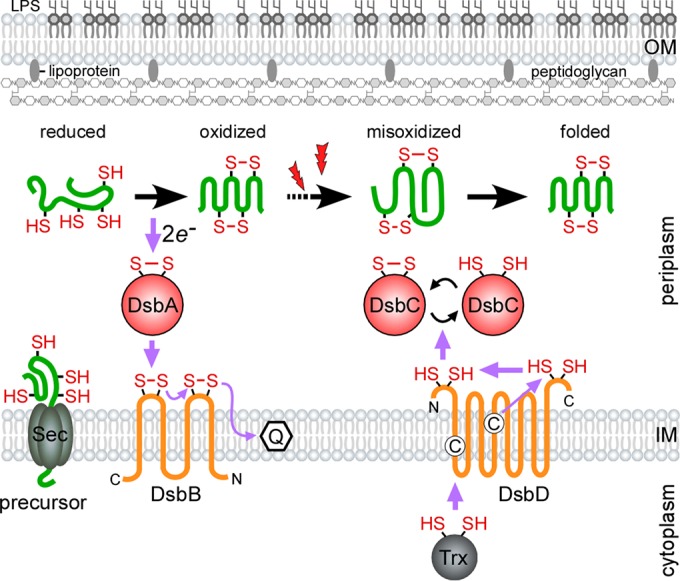
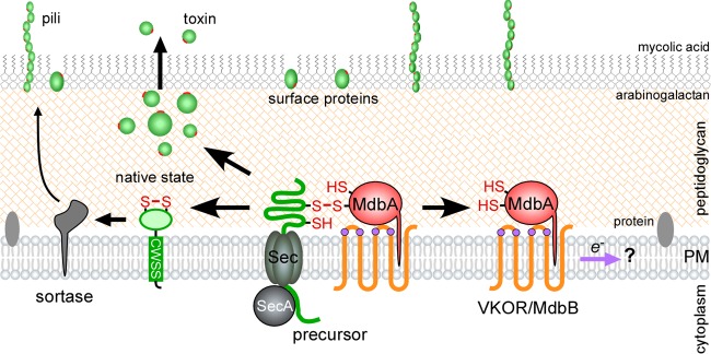
Disulfide bonds are important for the stability and function of many secreted proteins. In Gram-negative bacteria, these linkages are catalyzed by thiol-disulfide oxidoreductases (Dsb) in the periplasm. Protein oxidation has been well studied in these organisms, but it has not fully been explored in Gram-positive bacteria, which lack traditional periplasmic compartments. Recent bioinformatics analyses have suggested that the high-GC-content bacteria (i.e., actinobacteria) rely on disulfide-bond-forming pathways. In support of this, Dsb-like proteins have been identified in Mycobacterium tuberculosis, but their functions are not known. Actinomyces oris and Corynebacterium diphtheriae have recently emerged as models to study disulfide bond formation in actinobacteria. In both organisms, disulfide bonds are catalyzed by the membrane-bound oxidoreductase MdbA. Remarkably, unlike known Dsb proteins, MdbA is important for pathogenesis and growth, which makes it a potential target for new antibacterial drugs. This review will discuss disulfide-bond-forming pathways in bacteria, with a special focus on Gram-positive bacteria.


Figures

FIG 1
FIG 1
Oxidative protein folding in the Gram-negative periplasm. To catalyze oxidative protein folding, DsbA donates a reactive disulfide bond to reduced protein precursors as they are secreted into the oxidizing periplasm by SecYEG. Following catalysis, the DsbA active site is reoxidized by the transmembrane protein DsbB, which shuttles the gained electrons to the electron transport chain via a conjugated quinone. Extracellular oxidative stress (denoted by lightning bolts) or the lack of DsbA proofreading activity can cause substrates to become misoxidized. Aberrant disulfide bonds are reshuffled by the reductase DsbC. The reducing power of DsbC is maintained by the transmembrane DsbD, which receives electrons from cytoplasmic thioredoxin (adapted from reference 26). The purple arrows denote the direction of the electron flow, and the cysteine residues in the membrane domains of DsbD are shown as circled C's.
FIG 2
FIG 2
Oxidative protein folding in the actinobacterial exoplasm. Using pilus proteins and diphtheria toxin as model substrates, oxidative protein-folding pathways have been proposed in the actinobacterial pathogens A. oris and C. diphtheriae. Unfolded pilin precursors are oxidized by the membrane-tethered thiol-disulfide oxidoreductase MdbA. Proper folding is a prerequisite for sortase-mediated assembly of pili on the cell surface. A. oris MdbA is reoxidized by the transmembrane VKOR, while C. diphtheriae MdbA is hypothesized to be recycled by an unidentified factor called MdbB. It is not clear how VKOR or MdbB is reoxidized. The catalytic cysteine residues of VKOR are shown as purple circles, disulfide bonds formed in mature proteins are shown as red lines, and the purple arrow indicates the presumed direction of the electron flow (modified after references and 24).

References

    1. Anfinsen CB, Haber E, Sela M, White FH Jr. 1961. The kinetics of formation of native ribonuclease during oxidation of the reduced polypeptide chain. Proc Natl Acad Sci U S A 47:1309–1314. doi: 10.1073/pnas.47.9.1309. - DOI - PMC - PubMed
    1. Goldberger RF, Epstein CJ, Anfinsen CB. 1963. Acceleration of reactivation of reduced bovine pancreatic ribonuclease by a microsomal system from rat liver. J Biol Chem 238:628–635. - PubMed
    1. Givol D, Goldberger RF, Anfinsen CB. 1964. Oxidation and disulfide interchange in the reactivation of reduced ribonuclease. J Biol Chem 239:PC3114–PC3116. - PubMed
    1. Tian G, Xiang S, Noiva R, Lennarz WJ, Schindelin H. 2006. The crystal structure of yeast protein disulfide isomerase suggests cooperativity between its active sites. Cell 124:61–73. doi: 10.1016/j.cell.2005.10.044. - DOI - PubMed
    1. Creighton TE, Zapun A, Darby NJ. 1995. Mechanisms and catalysts of disulfide bond formation in proteins. Trends Biotechnol 13:18–23. doi: 10.1016/S0167-7799(00)88896-4. - DOI - PubMed

Publication types

MeSH terms

LinkOut - more resources