. 2016 Feb 5;291(6):2904-16.
doi: 10.1074/jbc.M115.690396. Epub 2015 Dec 7.

Binding of the Covalent Flavin Assembly Factor to the Flavoprotein Subunit of Complex II

Affiliations

Binding of the Covalent Flavin Assembly Factor to the Flavoprotein Subunit of Complex II

Elena Maklashina et al. J Biol Chem. .

Abstract

Escherichia coli harbors two highly conserved homologs of the essential mitochondrial respiratory complex II (succinate:ubiquinone oxidoreductase). Aerobically the bacterium synthesizes succinate:quinone reductase as part of its respiratory chain, whereas under microaerophilic conditions, the quinol:fumarate reductase can be utilized. All complex II enzymes harbor a covalently bound FAD co-factor that is essential for their ability to oxidize succinate. In eukaryotes and many bacteria, assembly of the covalent flavin linkage is facilitated by a small protein assembly factor, termed SdhE in E. coli. How SdhE assists with formation of the covalent flavin bond and how it binds the flavoprotein subunit of complex II remain unknown. Using photo-cross-linking, we report the interaction site between the flavoprotein of complex II and the SdhE assembly factor. These data indicate that SdhE binds to the flavoprotein between two independently folded domains and that this binding mode likely influences the interdomain orientation. In so doing, SdhE likely orients amino acid residues near the dicarboxylate and FAD binding site, which facilitates formation of the covalent flavin linkage. These studies identify how the conserved SdhE assembly factor and its homologs participate in complex II maturation.

Keywords: chaperone; complex II; flavin adenine dinucleotide (FAD); fumarate reductase; mitochondrial respiratory chain complex; protein assembly; protein self-assembly; protein-protein interaction; succinate dehydrogenase.


Figures

FIGURE 1.
FIGURE 1.
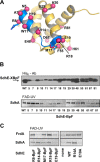
A, ribbon representation of SdhE (PDB file 1X6I, (25)). The surface amino acids SdhE-N5, -A7, -R8, -H10, -W11, -R14, -M17, -R18, -E19, -E29, -H30, -E48, -F55, -H61, -A67, and -R80 were individually substituted with photoactivatable BpF and are shown as red spheres. The conserved N-terminal region is shown in blue. The positions in SdhE where BpF was introduced and proved positive for cross-linking to FrdA are represented as sticks. B, expression levels of SdhE-BpF variants and covalent FAD levels in RP-3 cells co-transformed with pQE-SdhE/SdhA and pEVOL-pBpF. The top panel shows the level of SdhE-XBpF detected by His6 antibody (Ab) following induction of SdhE as described under “Experimental Procedures.” Whole cell lysates equal to 20 μg of protein were used for comparison. The arrow represents full-length SdhE-XBpF. Lanes on the right labeled 51, 61, 67, and 81 were from a different gel treated in an identical manner to those on the left. The lower panel shows the covalent FAD fluorescence of SdhA in the RP-3 cells as described under “Experimental Procedures.” Lanes labeled 29–81 on the right are from a different gel treated identically to those on the right. C, functional evaluation of SdhE-BpF variants for their ability to assist with covalent FAD incorporation into FrdA. Flavin fluorescence for the E. coli FrdA or SdhA subunits co-expressed with SdhE-XBpF variants in E. coli strain RP-3.
FIGURE 2.
FIGURE 2.
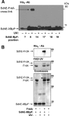
A, photo-cross-linking of SdhE-8BpF to FrdA. Cell lysates containing FrdA and individual SdhE-BpF variants at the indicated position were exposed to UV light for 90 min on ice (+) or left under ambient light (−) under identical conditions. SdhE was detected by Western blotting with anti-His6 antibodies. The ∼75-kDa band corresponds to the specific FrdA cross-linked product. B, cross-linking between purified SdhE-8BpF and FrdA. Purified holo-FrdA generated in RP-2 cells with the His tag cleaved with TEV protease was used for these studies. Isolated proteins were exposed to UV light for 45 min in 25 mm HEPES (pH 7.5). The top panel shows an immunoblot against the His6 tag of SdhE cross-linked to FrdA. The middle panel shows covalent FAD fluorescence where it is evident that the cross-linked product also contains covalent FAD. The bottom panel is the Coomassie Blue-stained gel. The asterisk indicates the cross-linked protein band that was excised from the SDS gel and characterized by mass spectral analysis. The ∼50-kDa protein that can be seen below native FrdA is a proteolytic fragment of FrdA.
FIGURE 3.
FIGURE 3.
Mass spectrometric identification of the photo-cross-linked residue in SdhE-R8BpF/FrdA. A, diagram of cross-linked peptides. BpF substituted amino acid represented as x. The presence of various b and y ions of different charge states within the spectrum is shown with the relative intensity of those fragments indicted by the color spectrum. B, full scan of predicted and actual isotopic peaks for cross-linked peptide. The stacked bar chart represents the predicted and observed proportions of the isotopic peaks. C, higher energy collisional dissociation spectrum with zoomed and annotated insets showing fragment ions corresponding to the cross-linked model shown in A.
FIGURE 4.
FIGURE 4.
FrdA and SdhA cross-linking to SdhE-R8BpF. A, ribbon diagram of flavoprotein subunit of complex II. Overlay of the E. coli SdhA subunit (PDB code 1NEK (4)) is shown in gray, and the FrdA subunit (PDB code 1KF6 (24)) is shown in cyan. The region where SdhE-R8BpF cross-links to the wild-type FrdA and SdhA-E186M is shown in the inset. B, SdhE-R8BpF cross-linking to FrdA and SdhA. Immunoblot of the 75-kDa region of SDS-PAGE gels with anti-His6 antibodies. Cell lysates containing SdhA and FrdA wild-type and variant proteins were incubated with lysates from cells harboring SdhE-R8BpF with (+) or without (−) exposure to UV light as indicated in Fig. 1C. C, FAD fluorescence of SdhA and FrdA variants. The covalent flavin fluorescence of WT and the SdhA and FrdA variants in cell lysates of RP-2 cells are shown. D, in vivo photo-cross-linking in RP-3 cells expressing His6-SdhE-R8BpF and the SdhA-E186M variant. Immunoblots using anti-His6 tag antibody shows the formation of the cross-linked product in cells that constitutively express SdhA-E186M and IPTG-induced His6-SdhE-R8BpF. The cells from three separate clones were exposed to UV light for 1 h prior to harvesting for immunoblot analysis. The lane with the molecular weight marker proteins between the UV exposed samples (+) and control (−) is removed for clarity because of the excessive signal of the marker proteins.
FIGURE 5.
FIGURE 5.
FrdA cross-linking to SdhE-M17BpF. A, molecular model showing juxtaposition of SdhE-R8 (gold sphere) and FrdA-M176 (cyan sphere). The panel on the right is rotated 90°. The FAD co-factor is shown as yellow sticks. B, residues in FrdA chosen for site-directed substitution to methionine. FrdA is positioned to show the proposed interacting surface with SdhE. The three amino acid residues proposed as potential sites for cross-linking to SdhE-BpF variants are shown as orange spheres. Also shown is the FrdA-M176 residue (cyan sphere) demonstrated to cross-link to SdhE-R8BpF. C, cross-linking of FrdA variants to SdhE-R8BpF and SdhE-M17BpF. Extracts from cells expressing wild-type FrdA or its variants were combined with purified SdhE-R8BpF or SdhE-M17BpF and exposed to UV light for 1 h as described in Fig. 2A. D, covalent FAD fluorescence of FrdA variants. The flavin fluorescence of FAD covalently bound to FrdA is shown from the experiment described in C. E, far UV CD spectra of purified SdhE-His6 WT enzyme, and the SdhE-R8BpF and SdhE-M17BpF variants. SdhE-WT enzyme data are shown as blue squares, SdhE-R8BpF data are shown as red triangles, and SdhE-M17BpF data are shown as black circles.
FIGURE 6.
FIGURE 6.
Mass spectrometric identification of the photo-cross-linked FrdA peptide to the SdhE-M17BpF variant. A, diagram of cross-linked peptides to FrdA-S239M. BpF substituted amino acid represented as x. The presence of various b and y ions of different charge states within the spectrum is shown with the relative intensity of those fragments indicted by the color spectrum. B, full scan of predicted and actual isotopic peaks for cross-linked peptide. The stacked bar chart represents the predicted and observed proportions of the isotopic peaks.
FIGURE 7.
FIGURE 7.
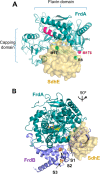
Model of SdhE interaction with the flavoprotein subunit of complex II based on two restraints. A, FrdA (PDB code 1KF6) is shown in teal with the regions that form cross-links to SdhE shown in magenta (FrdA-M176, the GLAAMEG peptide). SdhE (PDB code 1X6I) is shown as a surface representation with the amino acid residues forming cross-links to FrdA indicated as green spheres. The FAD co-factor in FrdA is shown as yellow sticks with the oxygen atoms in light red. The figure shows SdhE wedged between the capping and flavin domains to lock the dicarboxylate binding site of FrdA in its closed position. B, the structure of FrdA (teal) and FrdB (blue) (PDB code 1KF6) overlaid with modeled position of SdhE as shown in A. The FrdA and SdhE proteins are rotated 90° from what is shown in A. The modeling suggests that SdhE occupies a similar position to where the N-terminal domain of FrdB resides in assembled complex II. The three iron-sulfur clusters in FrdB (S1, [2Fe-2S]; S2, [4Fe-4S]; S3, [3Fe-4S]) are shown as stick representations, and the FAD co-factor in FrdA is shown as yellow sticks.
FIGURE 8.
FIGURE 8.
Structural comparison of SdhA with E. colil-aspartate oxidase. A, overlay of SdhA (PDB code 1NEK, shown in gray (4)) and LASPO in complex with FAD and succinate (PDB code 1KNP, shown in green (53)). The flavin and capping domains, as well as the helical domain of FrdA that interacts with SdhE, are labeled. B, comparison of LASPO structures. The apo form of LASPO is shown in magenta (PDB code 1CHU (54)), and LASPO in complex with FAD and succinate (PDB code 1KNP (53)) is shown in green. The position(s) of the capping domain in relation to the FAD domain are indicated by arrows.
FIGURE 9.
FIGURE 9.
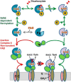
Scheme for SdhE function in covalent FAD incorporation into complex II. The apo-Fp is shown in the center of the scheme. Binding of FAD to the apo-Fp induces closure of the capping domain followed by interaction with SdhE, which assists in maintaining the closed conformation. Binding of a dicarboxylate at the active site in the presence of SdhE promotes the locked position of the active site domain, which facilitates the self-catalytic flavinylation reaction. The covalent FAD linkage is indicated by the blue vertical bar attached to FAD. Upon release of SdhE from holo-Fp, the protein forms a catalytic dimer with the iron-sulfur protein (Ip shown in green) that docks at the site previously occupied by SdhE. The Fp-Ip complex can then associate with the preformed membrane-spanning subunits to form functional complex II. Alternatively, in the absence of SdhE, association of apo-Fp in complex with FAD and Ip results in assembly of the membrane-bound complex, which is incapable of succinate oxidation (35). Once assembled with noncovalent FAD, the enzyme remains inactive. fum, fumarate; succ, succinate.

References

    1. Cecchini G. (2003) Function and structure of complex II of the respiratory chain. Annu. Rev. Biochem. 72, 77–109 - PubMed
    1. Iverson T. M., Luna-Chavez C., Cecchini G., and Rees D. C. (1999) Structure of the Escherichia coli fumarate reductase respiratory complex. Science 284, 1961–1966 - PubMed
    1. Lancaster C. R., Kröger A., Auer M., and Michel H. (1999) Structure of fumarate reductase from Wolinella succinogenes at 2.2 A resolution. Nature 402, 377–385 - PubMed
    1. Yankovskaya V., Horsefield R., Törnroth S., Luna-Chavez C., Miyoshi H., Léger C., Byrne B., Cecchini G., and Iwata S. (2003) Architecture of succinate dehydrogenase and reactive oxygen species generation. Science 299, 700–704 - PubMed
    1. Sun F., Huo X., Zhai Y., Wang A., Xu J., Su D., Bartlam M., and Rao Z. (2005) Crystal structure of mitochondrial respiratory membrane protein complex II. Cell 121, 1043–1057 - PubMed

Publication types

MeSH terms

Substances

LinkOut - more resources