. 2015 Dec 7;211(5):1077-91.
doi: 10.1083/jcb.201502036.

Ephrin-A3 promotes and maintains slow muscle fiber identity during postnatal development and reinnervation

Affiliations

Ephrin-A3 promotes and maintains slow muscle fiber identity during postnatal development and reinnervation

Danny A Stark et al. J Cell Biol. .

Abstract

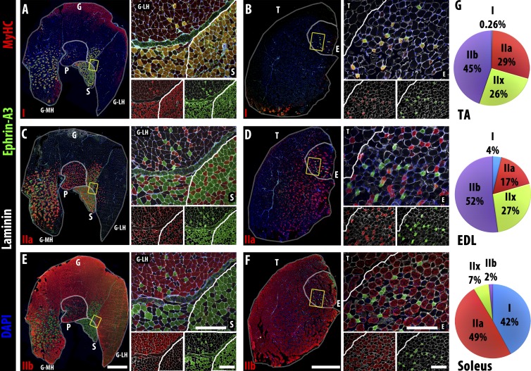
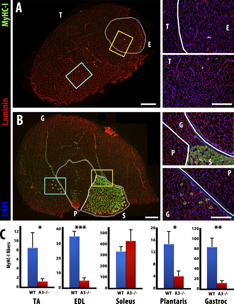
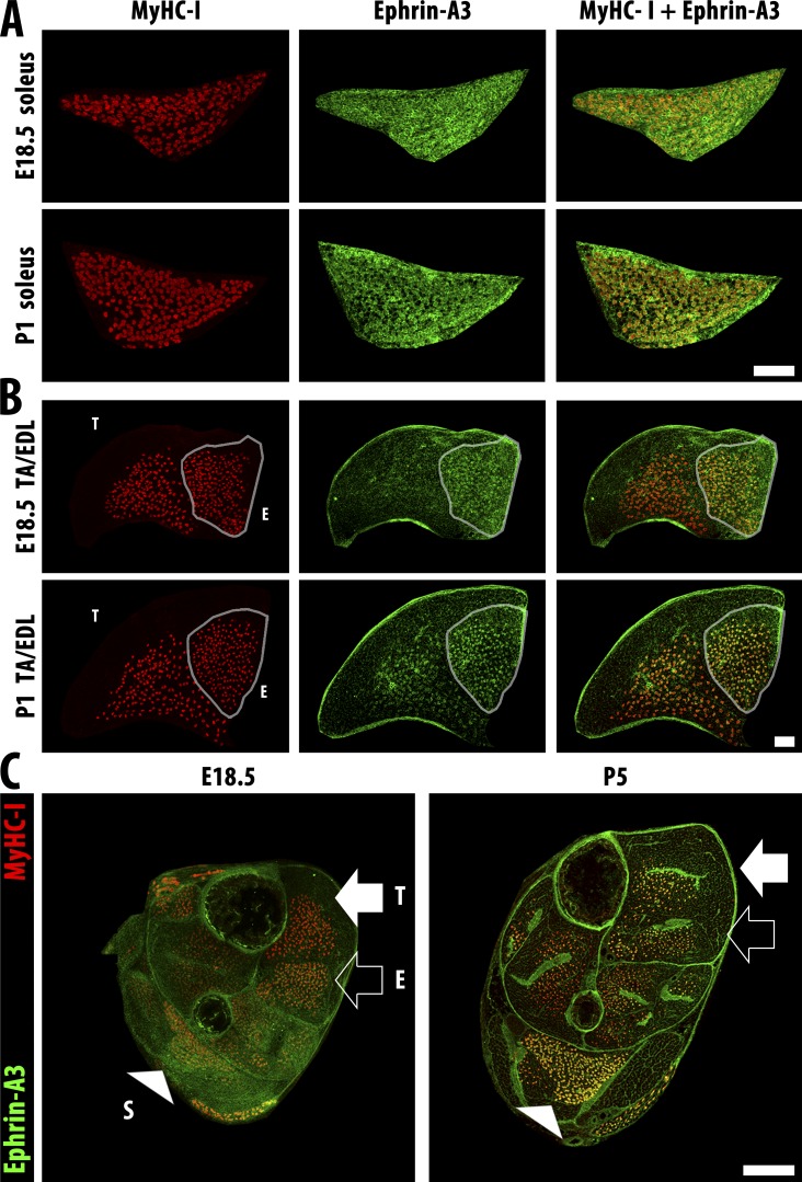
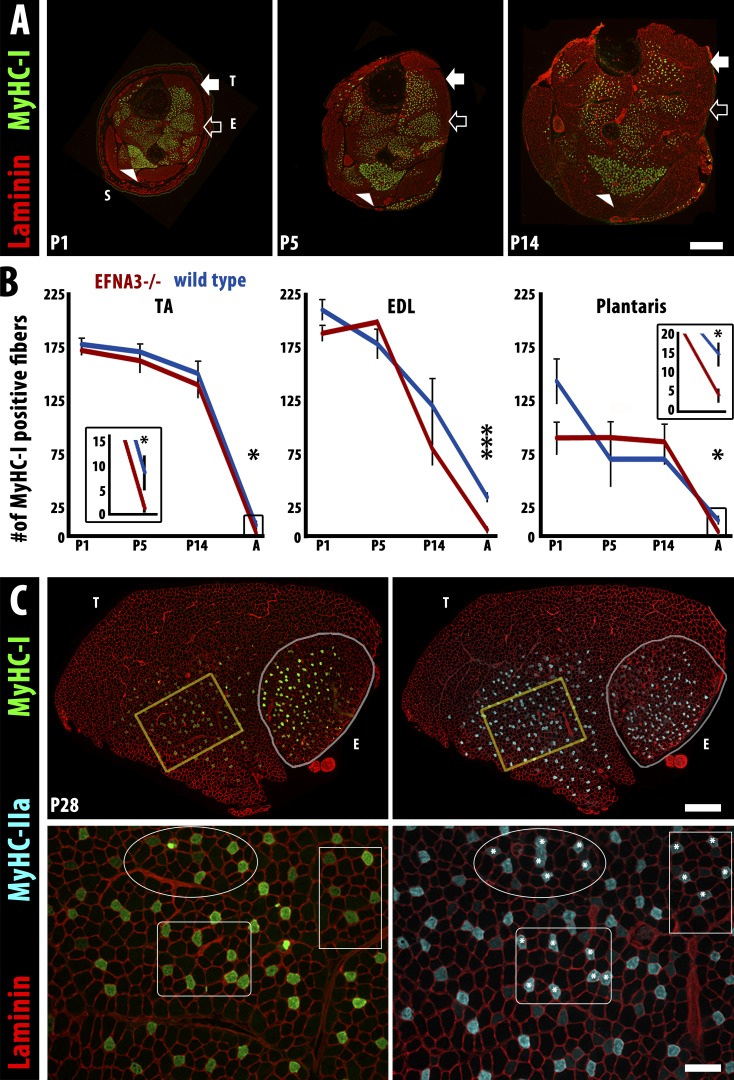
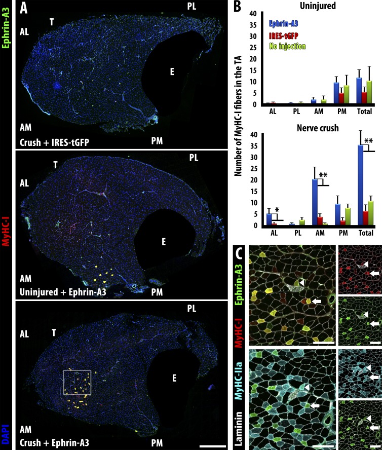
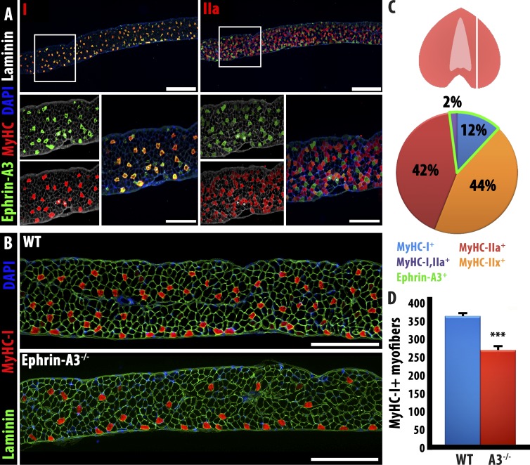
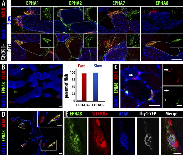
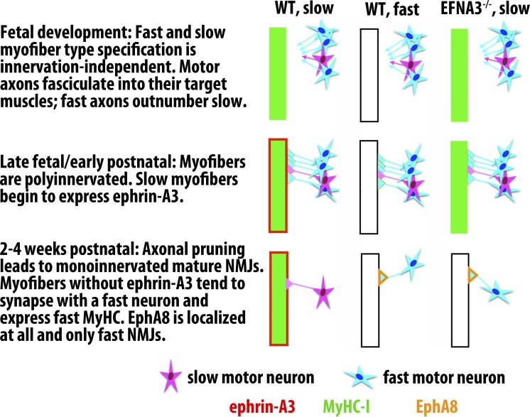
Each adult mammalian skeletal muscle has a unique complement of fast and slow myofibers, reflecting patterns established during development and reinforced via their innervation by fast and slow motor neurons. Existing data support a model of postnatal "matching" whereby predetermined myofiber type identity promotes pruning of inappropriate motor axons, but no molecular mechanism has yet been identified. We present evidence that fiber type-specific repulsive interactions inhibit innervation of slow myofibers by fast motor axons during both postnatal maturation of the neuromuscular junction and myofiber reinnervation after injury. The repulsive guidance ligand ephrin-A3 is expressed only on slow myofibers, whereas its candidate receptor, EphA8, localizes exclusively to fast motor endplates. Adult mice lacking ephrin-A3 have dramatically fewer slow myofibers in fast and mixed muscles, and misexpression of ephrin-A3 on fast myofibers followed by denervation/reinnervation promotes their respecification to a slow phenotype. We therefore conclude that Eph/ephrin interactions guide the fiber type specificity of neuromuscular interactions during development and adult life.


Figures

Figure 1.
Figure 1.
Ephrin-A3 is expressed on all MyHC-I+ve (slow) myofibers. (A–F) Immunohistochemistry showing localization of ephrin-A3 (green) compared with two of three fast MyHCs (red: MyHC-I, top row; MyHC-IIa, middle row; and MyHC-IIb, bottom row), along with nuclei (DAPI, blue) and laminin (white) on transverse cryosections from uninjured gastrocnemius (G), plantaris (P), soleus (S), TA (T), and EDL (E) muscles. The yellow boxes in the left of each panel indicate the magnified regions on the top right, which are separated underneath to show expression of MyHC/laminin (left) and ephrin-A3/laminin (right). (A and B) All slow, MyHC-I+ve myofibers in all muscles of the distal hindlimb express ephrin-A3. (C and D) No fast, MyHC-IIa+ve myofibers in the distal hindlimb express ephrin-A3. (E and F) Ephrin-A3 is also never expressed by fast, MyHC-IIb+ve myofibers. (G) Fiber type distribution of the TA, EDL, and soleus muscles (n = 3) in adult: each muscle has a unique and consistent ratio of the four myofiber types. Whole muscle montage gamma = 1.45. Bars: (montages) 1 mm; (insets) 300 µm. MH, gastrocnemius medial head; LH, lateral head.
Figure 2.
Figure 2.
Slow myofiber number is reduced in adult ephrin-A3−/− mice. (A) Immunohistochemistry showing laminin (red) and MyHC-I (green) expression in transverse cryosections from uninjured TA (T) and EDL (E) muscles of an adult ephrin-A3−/− mouse. (B) Immunohistochemistry showing laminin (red) and MyHC-I (green) expression in transverse cryosections from uninjured gastrocnemius (G), plantaris (P), and soleus (S) muscles of an adult ephrin-A3−/− mouse. Inset areas are enlarged at right (yellow, top; cyan, bottom). (C) All but one muscle scored had significantly fewer MyHC-I+ve myofibers in ephrin-A3−/− mice than age-matched wild type. P = 0.029 (TA); 0.0001 (EDL); 0.0035 (gastrocnemius); and 0.034 (plantaris). Error bars represent SEM. The change in MyHC-I+ve myofibers in the soleus is not significant. *, P < 0.05; **, P < 0.005; ***, P < 0.0005. Bars: (montages) 500 µm; (insets) 300 µm. n ≥ 3.
Figure 3.
Figure 3.
Slow fibers are specified embryonically and then express ephrin-A3 after birth. (A) Slow myofibers (MyHC-I, red) in the soleus are present at E18.5, but ephrin-A3 staining (green) remains diffuse until P1. (B) Slow myofibers (MyHC-I, red) in the EDL (outline) possess more specific ephrin-A3 (green) expression than the soleus at E18.5, but slow myofibers in the TA have only faint ephrin-A3 expression even at P1. Bars, 100 µm. (C) Transverse cryosections of distal hindlimbs of wild-type pups at E18.5 and P5 stained for ephrin-A3 (green) and MyHC-I (red) to highlight muscle-specific difference in coexpression pattern and intensity. The soleus (S, arrowhead), TA (T, closed arrow), and EDL (E, open arrow) muscles can be identified by the spatial pattern of MyHC-I+ve myofibers in each section. Bar, 500 µm.
Figure 4.
Figure 4.
The loss of slow myofibers in ephrin-A3−/− mice occurs postnatally by conversion to IIA fast myofibers. (A) Immunohistochemistry showing laminin (red) and MyHC-I (green) expression in transverse cryosections of distal hindlimbs of ephrin-A3−/− mice at P1, P5, and P14. The soleus (S, arrowhead), TA (T, closed arrow), and EDL (E, open arrow) muscles can be identified by the spatial pattern of MyHC-I+ve myofibers in each section. Bar, 500 µm. (B) Slow myofiber number in TA, EDL, and plantaris of wild-type and ephrin-A3−/− (EFNA3−/−) muscles are not significantly different until after at least 2 wk of postnatal life (insets show slow myofiber number in adult TA and plantaris at a different scale). Error bars = SEM. *, P < 0.05; ***, P < 0.001. (C) MyHC-I+ve myofibers in ephrin-A3−/− muscle sections at P28 (green, left) frequently coexpress MyHC-IIa (cyan, right), indicating that they are in transition to a faster fiber type; laminin (red) outlines myofibers on both sides. Insets (yellow boxes) are magnified under each panel: three regions are identified in each inset to facilitate comparison, and asterisks indicate MyHC-IIa+ve myofibers that are also MyHC-I+ve. Bars: (montages) 500 µm; (insets) 100 µm.
Figure 5.
Figure 5.
Misexpression of ephrin-A3 in the TA by electroporation followed by sciatic nerve crush imposes a fast to slow fiber type switch by 28 d postcrush. (A) Neither electroporation with a control plasmid (IRES-tGFP) followed by nerve crush (top) nor electroporation with ephrin-A3 plasmid without nerve crush (middle) significantly changed the number of slow myofibers in wild-type TA muscles compared with sham-operated controls. However, electroporation with ephrin-A3 plasmid followed by challenge with nerve crush led to significant conversion of fast myofibers to slow (MyHC-I+ve) at the injection site (bottom). EDL (E) is masked in all panels. Bar, 500 µm. (B) Quantification of slow myofibers in different quadrants of the TA in the experiments described above: AL, anterior lateral; AM, anterior medial; PL, posterior lateral; PM, posterior medial. Plasmid DNA was injected into the anterior aspect of the TA followed by electroporation. Only electroporation with ephrin-A3 in conjunction with nerve crush (blue bars, bottom) significantly increases MyHC-I+ve myofibers. Error bars = SEM. *, P < 0.05; **, P < 0.01. (C) Serial sections corresponding to inset box in A showing coexpression of ephrin-A3 (green) with either MyHC-I (red, top) or MyHC-IIa (cyan, bottom). Laminin is shown in white. All ephrin-A3+ve myofibers express MyHC-I (i.e., arrows); a few (arrowheads) express both MyHC-I and MyHC-IIa. Bars, 100 µm. T, TA; E, EDL.
Figure 6.
Figure 6.
Ephrin-A3−/− diaphragm muscle displays a phenotype intermediate between TA and soleus muscles. (A) As noted in hindlimb muscles, ephrin-A3 expression is detected on all MyHC-I+ve myofibers in the diaphragm; in sections taken as indicated in the cartoon at right, MyHC-I is expressed by 14% of myofibers scored, all of which also express ephrin-A3. Bars: (top) 400 µm; (bottom) 100 µm. (B) Equivalent sections of wild-type and ephrin-A3−/− adult diaphragm show a reduction, but not loss, of slow myofibers. Bars, 500 μm. (C) Representation of the plane of section in A and B and quantification of muscle fiber types and ephrin-A3 expression in WT diaphragm. (D) Quantification of Type I fibers through the same plane of section in wild-type and ephrin-A3−/− adult diaphragm; error bars represent SEM. WT, wild type. ***, P < 0.001.
Figure 7.
Figure 7.
EphA8 is expressed by terminal Schwann cells associated with fast myofiber NMJs. (A) EphA1, EphA2, and EphA7 (green) are present on NMJs (α-bungarotoxin, red) of both fast myofibers (gray outline) and slow myofibers (MyHC-I, blue), but EphA8 (green, right) is only detectable at NMJs of fast myofibers. Insets show green staining for the indicated Eph at the same magnification. Bar, 25 µm. (B) Section of soleus muscle (MyHC-I+ve myofibers, blue) showing expression of EphA8 (green) at fast but not slow myofiber NMJs (α-bungarotoxin, red). Bar, 25 µm. 100% of fast myofiber NMJs were positive for EphA8 (red bar), whereas almost all (97%) slow myofiber NMJs lacked EphA8 staining (blue bar); the very small fraction (<3%) of EphA8+ve NMJs associated with MyHC-I+ve myofibers may represent hybrid myofibers. n = 134 NMJs. (C) A fast TA myofiber converted to slow by ephrin-A3 misexpression/nerve crush does not have EphA8 at its NMJ: arrow indicates the NMJ of the same slow myofiber marked with an arrow in Fig. 5 C (all 13 converted myofibers scored were EphA8 negative). Bars, 25 µm. (D) EphA8 (green) expression is localized at the NMJ (α-bungarotoxin, red) and not associated with the motor axon (SMI-31, blue). Bars, 25 µm. α-Bungarotoxin gamma = 1.45. (E) Confocal analysis of staining for presynaptic neuron (Thy1-YFP, gray), postsynaptic myofiber (acetylcholine receptor, blue), and Schwann cell cytoplasm (S100b, red) consistent with expression of EphA8 (green) by terminal Schwann cells. Note that because S100b is cytoplasmic and EphA8 is at the cell surface, overlap of staining is not necessarily expected in these confocal sections. Bar, 10 µm.
Figure 8.
Figure 8.
Model for slow muscle fiber specification via ephrin-A3/EphA8 repulsive interactions during postnatal synaptic pruning. We hypothesize that after cell-autonomous slow myofiber specification during embryonic and fetal development, MyHC-I–expressing myofibers begin to express ephrin-A3, leading to preferential elimination of synapses with fast motor axons late in NMJ maturation. In the absence of ephrin-A3, because fast motor axons outnumber slow motor axons in most mixed fiber type muscles, this competitive advantage is lost and the slow myofiber population is largely lost as well because of innervation-dependent fiber type switching to a faster phenotype. It is not yet clear what lineage or local signaling factors induce a subset of terminal Schwann cells to become EphA8+ve, or when and how they become localized to fast NMJs, although they are present at birth and are restricted to maturing fast NMJs by P14 (Fig. S5). We therefore indicate expression of EphA8 only in the final, steady state. WT, wild type.

Similar articles

Cited by

References

    1. Agbulut O., Noirez P., Beaumont F., and Butler-Browne G.. 2003. Myosin heavy chain isoforms in postnatal muscle development of mice. Biol. Cell. 95:399–406. 10.1016/S0248-4900(03)00087-X - DOI - PubMed
    1. Aihara H., and Miyazaki J.. 1998. Gene transfer into muscle by electroporation in vivo. Nat. Biotechnol. 16:867–870. 10.1038/nbt0998-867 - DOI - PubMed
    1. Ashby P.R., Wilson S.J., and Harris A.J.. 1993. Formation of primary and secondary myotubes in aneural muscles in the mouse mutant peroneal muscular atrophy. Dev. Biol. 156:519–528. 10.1006/dbio.1993.1098 - DOI - PubMed
    1. Augusto V., Padovani C.R., and Campos G.E.R.. 2004. Skeletal muscle fiber types in C57BL6 mice. Braz. J. Morphol. Sci. 21:89–94.
    1. Ausoni S., Gorza L., Schiaffino S., Gundersen K., and Lømo T.. 1990. Expression of myosin heavy chain isoforms in stimulated fast and slow rat muscles. J. Neurosci. 10:153–160. - PMC - PubMed

Publication types

MeSH terms

LinkOut - more resources