. 2015 Dec 29;112(52):15940-5.
doi: 10.1073/pnas.1521818112. Epub 2015 Dec 7.

Molecular mechanisms underlying the exceptional adaptations of batoid fins

Affiliations

Molecular mechanisms underlying the exceptional adaptations of batoid fins

Tetsuya Nakamura et al. Proc Natl Acad Sci U S A. .

Abstract

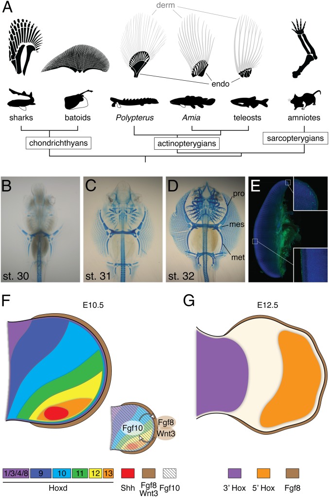
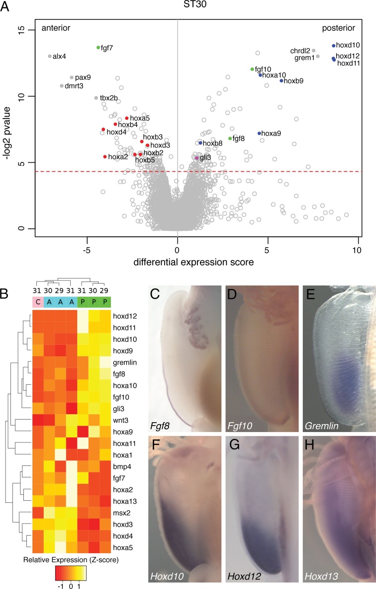
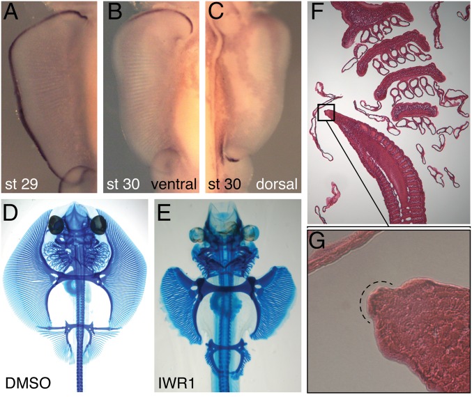
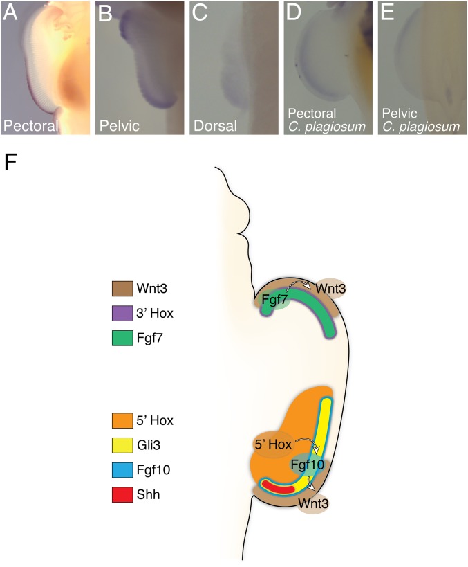
Extreme novelties in the shape and size of paired fins are exemplified by extinct and extant cartilaginous and bony fishes. Pectoral fins of skates and rays, such as the little skate (Batoid, Leucoraja erinacea), show a strikingly unique morphology where the pectoral fin extends anteriorly to ultimately fuse with the head. This results in a morphology that essentially surrounds the body and is associated with the evolution of novel swimming mechanisms in the group. In an approach that extends from RNA sequencing to in situ hybridization to functional assays, we show that anterior and posterior portions of the pectoral fin have different genetic underpinnings: canonical genes of appendage development control posterior fin development via an apical ectodermal ridge (AER), whereas an alternative Homeobox (Hox)-Fibroblast growth factor (Fgf)-Wingless type MMTV integration site family (Wnt) genetic module in the anterior region creates an AER-like structure that drives anterior fin expansion. Finally, we show that GLI family zinc finger 3 (Gli3), which is an anterior repressor of tetrapod digits, is expressed in the posterior half of the pectoral fin of skate, shark, and zebrafish but in the anterior side of the pelvic fin. Taken together, these data point to both highly derived and deeply ancestral patterns of gene expression in skate pectoral fins, shedding light on the molecular mechanisms behind the evolution of novel fin morphologies.

Keywords: AER; development; evolution; fin; skate.


Conflict of interest statement

The authors declare no conflict of interest.

Figures

Fig. 1.
Fig. 1.
Appendage diversity and the skate unique fin. (A) The pectoral fin and forelimb skeleton in a variety of taxa. Chondrichthyan and basal actinopterygian fins are composed of three bones, pro-, meso-, and metapterygium, whereas the sarcopterygian appendage is a single rod of the metapterygium axis. The batoid fin is extremely wide along the A–P axis compared with other vertebrates. Dermal bones (derm) and endochondral bones (endo) are labeled by gray and black colors depending on the difference of its developmental mechanisms (36). (B–D) Alcian Blue-stained skeletal preparations of skate embryos at stages 30–32. mes, mesopterygium; met, metapterygium; pro, propterygium. Both pectoral and pelvic fins elongate along the A–P axis. (E) Immunostaining for phosphorylated histone H3 (green) and DAPI (blue) in the pectoral fin at stage 31. Image composed using tiled scanning by confocal microscope (Zeiss ZEN software). Inset, magnified portions of the anterior and central fin. Statistical analysis of cell proliferation rates in each portion can be found in SI Appendix, Fig. S1. (F and G) Summary of the developmental mechanisms of the tetrapod limb. At an early stage (F), Shh is expressed in the posterior limb bud, and 5′Hox genes show a gradient of expression. Fgf10 induces and maintains AER structure. In turn, Fgf8 and Wnt3 in AER stimulate cell proliferation in the limb mesenchyme. As the limb bud develops, 5′Hox genes mark the autopod domain, whereas 3′Hox genes are expressed in the proximal limb.
Fig. 2.
Fig. 2.
Analysis of skate fin development by RNA-seq. (A) Differential expression of transcripts assembled from RNA-seq of anterior and posterior fin tissue specimens of stage 30 skates. Differential expression score is derived from multiple A–P comparisons of transcript abundance (n = 3; see SI Appendix). Above the red dotted line corresponds to P values less than 0.05. Blue, 5′Hox genes; green, Fgf genes; purple, Gli3; red, 3′Hox genes. (B) Standardized read counts (z-scores) for selected transcripts in the pectoral fin at three developmental stages and locations of the pectoral fin. Transcripts were median-summarized according to their annotations. A, anterior fin; C, center fin; P, posterior fin. Sample developmental stages are listed at the top, and clustering is based on relative expression levels. (C–H) Whole-mount in situ hybridization for canonical tool kit genes for limb development at stage 30. (C) Fgf8. The expression is at the posterior ectoderm. (D) Fgf10. The expression can be seen at the posterior mesenchyme. (E) Gremlin. (F) Hoxd10. (G) Hoxd12. (H) Hoxd13. Note that all expression patterns are limited to the posterior half, not in the anterior fin.
Fig. 3.
Fig. 3.
Dual AERs in the developing paired fins of skates. (A–C) Wnt3 expression in the pectoral fin of skate at stage 29 (A) and 30 (B, ventral view; C, dorsal view). Wnt3 expression is a continuous strip in the ectoderm at stage 29, but splits into the anterior and posterior domain after stage 30. (D and E) Alcian Blue-stained skeletal preparations of skate embryos treated by DMSO or the WNT inhibitor IWR1. Pro- and mesopterygium were affected and shorter in IWR1-treated embryos compared with the control embryo. (F and G) Sagittal section of skate embryo at stage 30. An AER-like structure can be observed at the anterior tip of the pectoral fin (G).
Fig. 4.
Fig. 4.
An ancient genetic module underlies the pro- and mesopterygium. (A–D) The 3′Hox gene expression patterns at stage 30. Each gene is expressed in the anterior mesenchyme. The expression domain is limited to only a narrow strip underlying the ectoderm. (E) Hoxa5 in the pectoral fin of the shark (C. plagiosum), where expression is in the anterior and posterior fin mesenchyme. (F and G) Fgf7 expression at stage 30 and 31. (H and I) Wnt3 expression 1 d after the implantation of DMSO (H) or FGF7 beads (I). (J and K) Wnt3 in situ hybridization in the pectoral fin at stage 31 following DMSO (J) or SU5402 (K) injection into the anterior fin. (L and M) Alcian Blue staining following DMSO or SU5402 injection into the anterior fin. The tip of the pectoral fin regressed in SU5402 injection (arrowheads).
Fig. 5.
Fig. 5.
Gli3 expression inversion and the mechanism of skate fin development. (A–C) Gli3 expression at stage 30 in the pectoral (A), pelvic (B), and dorsal fin (C) of skate. Gli3 is expressed in the posterior side of the pectoral and dorsal fin while it is expressed in the anterior pelvic fin. The expression in the posterior tip of pelvic fin is likely in the clasper. (D and E) Gli3 expression at stage 30 in the pectoral (D) or pelvic fin (E) of shark. Gli3 is expressed in the posterior pectoral fin and in the anterior pelvic fin, as seen in skate. (F) Summary of molecular mechanisms controlling skate unique fin development. Although the posterior genetic module is similar to that of tetrapods, the anterior genetic module is distinct. Fgf7 and 3′Hox genes are expressed in the anterior mesenchyme and induce Wnt3 expression, resulting in an extra AER-like tissue in the anterior fin. The posterior AER and anterior AER-like tissue extend the fin in posterior and anterior directions. Note that Gli3 is coexpressed with Shh and 5′Hox in the posterior fin.

References

    1. Alfred Sherwood Romer . The Vertebrate Body. WB Saunders; Philadelphia: 1949.
    1. Nelson JS. Fishes of the World. 4th Ed Wiley; New Jersey: 2006.
    1. Janvier P. Early Vertebrates. Clarendon Press; Oxford: 1996.
    1. Aschliman NC, et al. Body plan convergence in the evolution of skates and rays (Chondrichthyes: Batoidea) Mol Phylogenet Evol. 2012;63(1):28–42. - PubMed
    1. Maxwell EE, Fröbisch NB, Heppleston AC. Variability and conservation in late chondrichthyan development: Ontogeny of the winter skate (Leucoraja ocellata) Anat Rec (Hoboken) 2008;291(9):1079–1087. - PubMed

Publication types

MeSH terms

Associated data

LinkOut - more resources