. 2016 Apr 30;22(2):333-43.
doi: 10.5056/jnm15127.

Acute Effects of Transforming Growth Factor-β1 on Neuronal Excitability and Involvement in the Pain of Rats with Chronic Pancreatitis

Affiliations

Acute Effects of Transforming Growth Factor-β1 on Neuronal Excitability and Involvement in the Pain of Rats with Chronic Pancreatitis

Xiaoyu Zhang et al. J Neurogastroenterol Motil. .

Abstract

Background/aims: This study was to investigate whether transforming growth factor-β1 (TGF-β1) plays a role in hyperalgesia in chronic pancreatitis (CP) and the underlying mechanisms.

Methods: CP was induced in male adult rats by intraductal injection of trinitrobenzene sulfonic acid (TNBS). Abdominal hyperalgesia was assessed by referred somatic behaviors to mechanical stimulation of rat abdomen. Dil dye injected into the pancreas was used to label pancreas-specific dorsal root ganglion (DRG) neurons. Whole cell patch clamp recordings and calcium imaging were performed to examine the effect of TGF-β1 on acutely isolated pancreas-specific DRG neurons. Western blot analysis was carried out to measure the expression of TGF-β1 and its receptors.

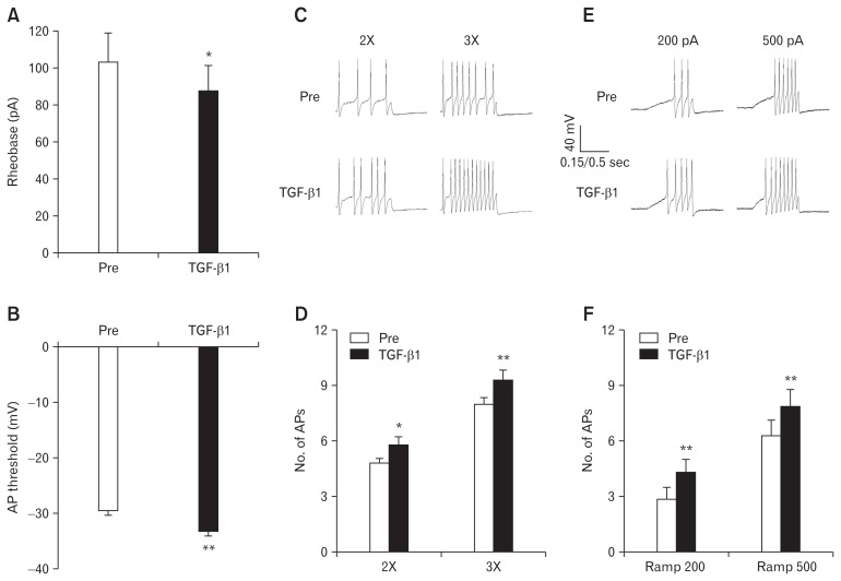
Results: TNBS injection significantly upregulated expression of TGF-β1 in the pancreas and DRGs, and TGF-β1 receptors in DRGs (T9-T13)in CP rats. Intrathecal injection of TGF-β receptor I antagonist SB431542 attenuated abdominal hyperalgesia in CP rats. TGF-β1 application depolarized the membrane potential and caused firing activity of DRG neurons. TGF-β1 application also reduced rheobase, hyperpolarized action potential threshold, and increased numbers of action potentials evoked by current injection of pancreas-specific DRG neurons. TGF-β1 application also increased the concentration of intracellular calcium of DRG neurons, which was inhibited by SB431542. Furthermore, intrathecal injection of TGF-β1 produced abdominal hyperalgesia in healthy rats.

Conclusions: These results suggest that TGF-β1 enhances neuronal excitability and increases the concentration of intracellular calcium. TGF-β1 and its receptors are involved in abdominal hyperalgesia in CP. This and future study might identify a potentially novel target for the treatment of abdominal pain in CP.

Keywords: Abdominal pain; Chronic pancreatitis; Dorsal root ganglion; Transforming growth factor beta 1.


Figures

Figure 1
Figure 1
Trinitrobenzene sulfonic acid (TNBS) injection up-regulates expression of transforming growth factor-β1 (TGF-β1) and TGF-β receptors. (A, B) TNBS injection significantly enhanced the expression of TGF-β1 and latent TGF-β1 complex in the pancreas of rats (*P < 0.05, n = 4 per group, two-sample t test). (C–F) TNBS injection significantly enhanced the expression of TGF-β receptor I and II, TGF-β1, and latent TGF-β1 complex in the pancreatic dorsal root ganglions (DRGs) of rats (*P < 0.05, n = 4 per group, two-sample t test). CON, control.
Figure 2
Figure 2
Transforming growth factor-β (TGF-β) receptor I antagonist SB431542 attenuates mechanical hyperalgesia. (A) Intrathecal injection (i.t.) of SB431542 significantly reduced response frequency of chronic pancreatitis (CP) rats to von Frey filament (VFF) stimuli (10 g) (**P < 0.01, ***P < 0.001 versus normal saline [NS]; n = 7 per group). (B) SB431542 at 100 μg/kg (i.t.) had no significant effect on response frequency of control rats (n = 7). (C) Daily intrathecal injection of SB431542 at 100 μg/kg for 7 days significantly reduced the response frequency of CP rats, which last for 24 hours (**P < 0.01, ***P < 0.001 versus Pre [before administration]; n = 7). (D) SB431542 at 100 μg/kg (i.t.) did not produce any effect on time for the CP rats to stay on the rotation bar (n = 8). Paired sample t test (B–D), two-way ANOVA followed by Tukey post hoc test (A).
Figure 3
Figure 3
Transforming growth factor-β1 (TGF-β1) produces mechanical hyperalgesia. (A) TGF-β1 (intrathecal injection [i.t.]) produced an increase of response frequency of control rats to von Frey filament (VFF) stimuli (10 g) (*P < 0.05, ***P < 0.001 versus normal saline [NS]; n = 7 per group; two-way ANOVA followed by Tukey post hoc test). (B) TGF-β1 at 10 ng per rat (i.t.) did not produce any effect on time for the control rats to stay on the rotation bar (n = 8, paired sample t test).
Figure 4
Figure 4
Transforming growth factor-β1 (TGF-β1) induces depolarization and firing activity of dorsal root ganglion (DRG) neurons. (A) DiI-fluorescence (left) and bright-field (right) images of acutely isolated DRG neurons. Pancreas-specific neurons are shown in red in DiI-fluorescence image. Bar = 25 μm. (B) The typical current-clamp recordings of DRG neurons showing firings, depolarization or no response after application of 10 ng/mL TGF-β1. (C) The percentage of DRG neurons showing firings (28.6%), depolarization (33.3%), or no response (38.1%) after application of TGF-β1 (n = 55 neurons in total). (D) Resting membrane potentials (RPs) before TGF-β1 application (Pre), peak value of RPs during TGF-β1 application (TGF-β1) and RPs after washout (Washout) (**P < 0.01 for TGF- β1 versus Pre, and Washout versus TGF-β1; n = 18; one-way repeated measures ANOVA followed by Tukey post hoc test).
Figure 5
Figure 5
Transforming growth factor-β1 (TGF-β1) enhances excitability of dorsal root ganglion neurons. (A) TGF-β1 application significantly reduced rheobase (*P < 0.05, n = 19). (B) TGF-β1 application markedly decreased action potential (AP) threshold (**P < 0.01, n = 19). (C) Typical traces of APs evoked by 2 times (2×) and 3 times (3×) rheobase current stimulation. (D) Bar graph shows the numbers of APs evoked by 2× and 3× rheobase current stimulation were significantly increased after TGF-β1 application (*P < 0.05, **P < 0.01; n = 19). (E) Typical traces of APs evoked by 200 pA and 500 pA ramp current stimulation. (F) Bar graph shows the numbers of AP evoked by 200 pA and 500 pA ramp current stimulation were significantly increased after TGF-β1 application (**P < 0.01, n = 19). Paired sample t test (A–F). Pre, before TGF-β1 application.
Figure 6
Figure 6
Transforming growth factor-β1 (TGF-β1) increases intracellular calcium ion concentration ([Ca2+]i) via TGF-β receptor I. (A) Representative traces of changes in [Ca2+]i (R(340/380)) in dorsal root ganglion neurons induced by 10 ng/mL TGF-β1 (top) and bar graph (bottom) shows no desensitization of TGF-β1-evokded [Ca2+]i increase after the second time application with an interval of 10 minutes (n = 32). (B) Representative traces of the inhibitory effect of TGF-β receptor I antagonist, SB431542, at the dose of 10 μM on TGF-β1-evoked [Ca2+]i increase. Bar graph shows SB431542 inhibited 80% of TGF-β1-evoked [Ca2+]i increase, which was partly reversed after washout (***P < 0.001 versus Pre, *P < 0.05 versus SB431542; n = 43). (C) Representative traces (top) and bar graph (bottom) show capsazepine (CZP) at the dose of 10 μM did not alter the TGF-β1-evoked [Ca2+]i increase (n = 28). One-way repeated measures ANOVA followed by Tukey post hoc test (A–C). Pre, before SB431542 or CZP application; Washout, after washout.

References

    1. Warshaw AL, Banks PA, Fernandez-Del Castillo C. AGA technical review: treatment of pain in chronic pancreatitis. Gastroenterology. 1998;115:765–776. doi: 10.1016/S0016-5085(98)70157-X. - DOI - PubMed
    1. Basbaum AI, Woolf CJ. Pain. Curr Biol. 1999;9:R429–R431. doi: 10.1016/S0960-9822(99)80273-5. - DOI - PubMed
    1. Xu GY, Winston JH, Shenoy M, Yin H, Pasricha PJ. Enhanced excitability and suppression of A-type K+ current of pancreas-specific afferent neurons in a rat model of chronic pancreatitis. Am J Physiol Gastrointest Liver Physiol. 2006;291:G424–G431. doi: 10.1152/ajpgi.00560.2005. - DOI - PubMed
    1. Xu GY, Winston JH, Shenoy M, Yin H, Pendyala S, Pasricha PJ. Transient receptor potential vanilloid 1 mediates hyperalgesia and is up-regulated in rats with chronic pancreatitis. Gastroenterology. 2007;133:1282–1292. doi: 10.1053/j.gastro.2007.06.015. - DOI - PubMed
    1. Cheng JK, Ji RR. Intracellular signaling in primary sensory neurons and persistent pain. Neurochem Res. 2008;33:1970–1978. doi: 10.1007/s11064-008-9711-z. - DOI - PMC - PubMed

LinkOut - more resources