. 2016 Apr;157(4):806-817.
doi: 10.1097/j.pain.0000000000000439.

Toll-like receptor 4 contributes to chronic itch, alloknesis, and spinal astrocyte activation in male mice

Affiliations

Toll-like receptor 4 contributes to chronic itch, alloknesis, and spinal astrocyte activation in male mice

Tong Liu et al. Pain. 2016 Apr.

Abstract

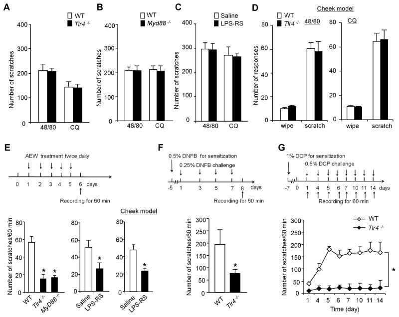
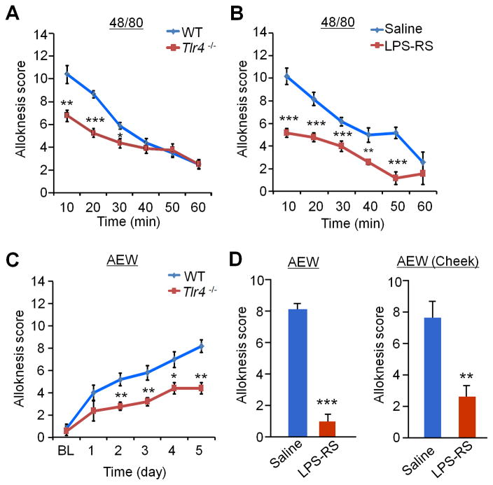
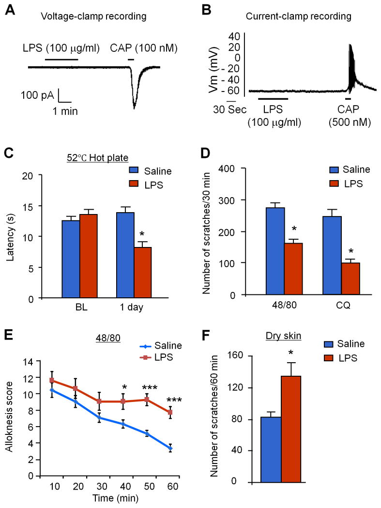
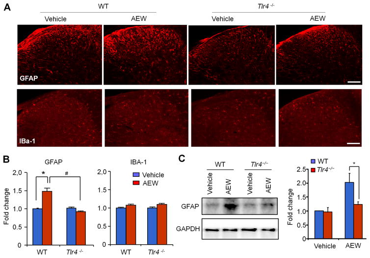
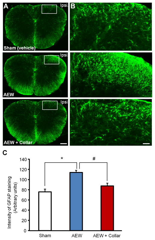
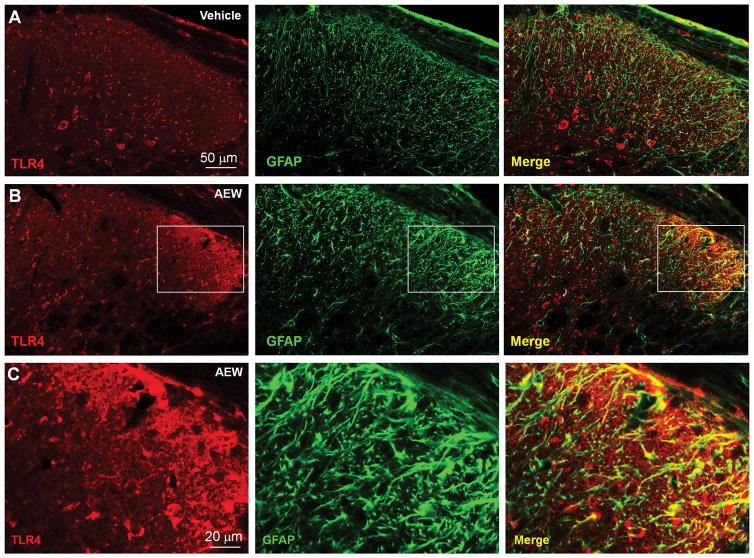
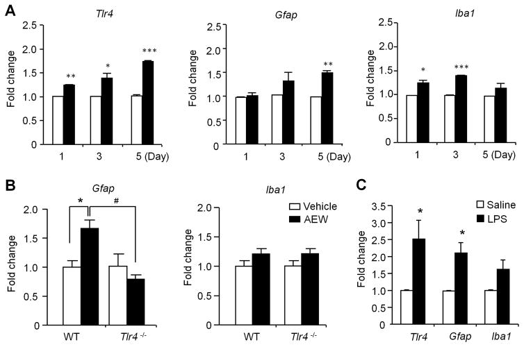
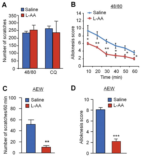
Increasing evidence suggests that Toll-like receptor 4 (TLR4) contributes importantly to spinal cord glial activation and chronic pain sensitization; however, its unique role in acute and chronic itch is unclear. In this study, we investigated the involvement of TLR4 in acute and chronic itch models in male mice using both transgenic and pharmacological approaches. Tlr4 mice exhibited normal acute itch induced by compound 48/80 and chloroquine, but these mice showed substantial reductions in scratching in chronic itch models of dry skin, induced by acetone and diethylether followed by water (AEW), contact dermatitis, and allergic contact dermatitis on the neck. Intrathecal (spinal) inhibition of TLR4 with lipopolysaccharide Rhodobacter sphaeroides did not affect acute itch but suppressed AEW-induced chronic itch. Compound 48/80 and AEW also produced robust alloknesis, a touch-elicited itch in wild-type mice, which was suppressed by intrathecal lipopolysaccharide R sphaeroides and Tlr4 deletion. Acetone and diethylether followed by water induced persistent upregulation of Tlr4 mRNA and increased TLR4 expression in GFAP-expressing astrocytes in spinal cord dorsal horn. Acetone and diethylether followed by water also induced TLR4-dependent astrogliosis (GFAP upregulation) in spinal cord. Intrathecal injection of astroglial inhibitor L-α-aminoadipate reduced AEW-induced chronic itch and alloknesis without affecting acute itch. Spinal TLR4 was also necessary for AEW-induced chronic itch in the cheek model. Interestingly, scratching plays an essential role in spinal astrogliosis because AEW-induced astrogliosis was abrogated by putting Elizabethan collars on the neck to prevent scratching the itchy skin. Our findings suggest that spinal TLR4 signaling is important for spinal astrocyte activation and astrogliosis that may underlie alloknesis and chronic itch.


Conflict of interest statement

Conflict of interest

All the authors have no competing financial interest in this study.

Figures

Figure 1
Figure 1. TLR4 signaling is required for chronic itch but not acute itch in the back and cheek models
(A, B) Acute itch induced by intradermal injection on the back skin of compound 48/80 (48/80, 100 μg) and chloroquine (CQ, 200 μg) is not altered in Tlr4−/− mice (A) and Myd88−/− mice (B). n = 5 mice per group. (C) Intrathecal LPS-RS (20 μg) does not affect 48/80 and CQ-induced acute itch. n = 5 mice per group. (D) Acute itch and pain in mouse cheek model. Note that intradermal injection of 48/80 or CQ induced comparable wiping and scratching in WT and Tlr4−/− mice. n=5–7 mice per group. (E) Dry skin after treatment with AEW (acetone and diethyether followed by water) induces chronic itch, which is substantially reduced in Tlr4−/− mice and Myd88−/− mice and also suppressed by intrathecal LPS-RS. Top, paradigm of AEW treatment. Low right, intrathecal LPS-RS administered 5 d after AEW treatment in either neck model or cheek model suppressed AEW-evoked scratching on day 6. *P< 0.05, compared with WT mice or corresponding vehicle groups, n = 5–7 mice per group. (F–G) Chronic itch in the neck models after contact allergic dermatitis, evoked by 2,4-dinitrofluorobenzene (DNFB, F), and atopic dermatitis, evoked by diphenylcyclopropenone (DCP, G) is substantially reduced in Tlr4−/− mice. Top panels, paradigms of DNFB and DCP treatment. *P<0.05, compared with WT control mice, student t-test (F) and Two-way ANOVA (G); n = 5–6 mice per group. Note that the neck models were tested for all the experimental conditions but the cheek models were only used for some conditions and specifically indicated in each graph. Data are presented as means ± S.E.M.
Figure 2
Figure 2. Alloknesis under both acute and chronic itch conditions is impaired in Tlr4−/− mice and suppressed by intrathecal LPS-RS in the back and cheek models
(A) Alloknesis, induced 30 min after compound 48/80 (48/80) injection, is partially reduced in Tlr4−/− mice. (B) Alloknesis, induced 30 min after 48/80 injection, is suppressed by intrathecal injection of LPS-RS (20 μg). (C) Alloknesis, induced after AEW-induced dry skin, is partially reduced in Tlr4−/− mice. (D) AEW-induced alloknesis at Day 6 in either neck model or cheek model is suppressed by intrathecal LPS-RS (20 μg). Data are presented as means ± S.E.M. *P<0.05, **P<0.01, ***P<0.001, compared with WT mice or saline vehicle group, Student’s t test; n = 5–6 mice per group.
Figure 3
Figure 3. LPS has no direct effects on neuronal excitability in cultured DRG neurons after bath application but enhances acute pain and chronic itch and suppresses acute itch in the back model after intrathecal injection
(A, B) LPS (100 μg/ml) fails to induce inward currents (A, n=25 neurons) and action potentials (B, n=8 neurons) in dissociated small-sized DRG neurons. Note that capsaicin (100 nM) induces inward currents and action potentials in these neurons. (C) Intrathecal LPS (10 μg) induces heat hyperalgesia 1 day after the injection. BL, baseline. (D) Intrathecal LPS (10 μg) suppresses 48/80 and CQ-induced acute itch. (E) Intrathecal LPS (10 μg) increases compound 48/80-induced alloknesis. (F) Intrathecal LPS (10 μg) potentiates AEW-induced chronic itch. Data are presented as means ± S.E.M. *P< 0.05, **P< 0.01, ***P< 0.001, compared with vehicle group, Student’s t test; n = 6–8 mice per group.
Figure 4
Figure 4. AEW treatment for 5 days in the back model induces astrogliosis but not microgliosis in cervical spinal cord dorsal horn (C3–C4) via TLR4
(A) Immunohistochemical staining showing the expression of astrogliosis marker GFAP and microgliosis marker IBA-1 following AEW treatment for 5 Days in WT and Tlr4−/− mice. Scale bars, 100 μm. (B) Quantitative analysis of GFAP and IBA-1 immuofluorescence intensity (as fold of WT vehicle control) in spinal cord dorsal horn of WT and Tlr4−/− mice 5 days after AEW treatment. *P<0.05; #P<0.05, n = 5 mice per group. (C) Western blot analysis of GFAP expression in spinal cord dorsal horn of WT and Tlr4−/− mice 5 days after AEW treatment. Right, quantitative analysis of GFAP western band intensity (as fold of WT control, normalized to GAPDH). *P<0.05, Student’s t test; n = 5 mice per group.
Figure 5
Figure 5. Scratching the dry skin is necessary for spinal cord astrogliosis in the cheek model
(A) Representative pictures of immunohistochemical staining showing the expression of astrogliosis marker GFAP in C1–C2 cervical spinal cord following AEW treatment for 5 days in cheek skin. Scale, 100 μm. (B) Enlarged images in the boxes of (A). Scale, 50 μm. (C) Quantitative analysis of GFAP immuofluorescence intensity (Arbitrary Units) in spinal cord dorsal horn of CD-1 mice 5 days after AEW treatment. Note that AEW-induced GFAP expression in the C1–C2 cervical spinal cord following AEW treatment was blocked by mouse wearing of an Elizabethan Collar that can prevent mouse from scratching the itchy skin. *P<0.05; #P<0.05, compared with corresponding group; Student’s t test; n = 4 mice per group.
Figure 6
Figure 6. AEW treatment for 5 days in the back model increases TLR4 expression in spinal cord astrocytes
(A,B) Double immunostaining of TLR4 with GFAP in the spinal cord dorsal horn (C3–C4) of vehicle (A) and AEW (5 d)-treated mice. Note that AEW increases GFAP-IR and TLR4-IR. Also note there is much more GFAP/TLR4 co-localization in chronic itch. Scale bar, 50 μm. (C) High magnification images from the boxes in B. Scale bars, 20 μm.
Figure 7
Figure 7. Quantitative RT-PCR shows the expression of Tlr4 and glia-related genes in cervical spinal cord dorsal horn (C3–C4) after AEW treatment on the back skin or intrathecal LPS injection
(A) Relative expression levels of Tlr4, Gfap, and Iba1 1, 3, and 5 days after AEW treatment. *P<0.05, **P<0.01, **P<0.001, compared with corresponding vehicle control, n = 4 mice per group. (B) Relative expression levels of Gfap and Iba1 in the dorsal horn of WT and Tlr4−/− mice 5 d after AEW treatment. *P<0.05; #P<0.05, n = 5 mice per group. Note that dry skin induces Tlr4-dependent Gfap expression. (C) Relative expression of Tlr4, Gfap, and Iba1 in cervical spinal cord dorsal horn 24 h after intrathecal LPS (10 μg) injection. Data are presented as means ± S.E.M. *P<0.05, compared with corresponding vehicle control, Student’s t test; n = 4 mice per group.
Figure 8
Figure 8. Intrathecal astroglial inhibitor L-alpha aminoadipate (L-AA, 100 nmol) reduces chronic itch and alloknesis without affecting acute itch
(A) Acute itch induced by compound 48/80 (48/80, 100 μg) and chloroquine (CQ, 200 μg) is not affected intrathecal L-AA. n = 6 mice per group. (B) 48/80-induced alloknesis is inhibited by intrathecal L-AA. *P<0.05, **P<0.01, compared with saline control, n = 6 mice per group. (C–D) AEW-induced chronic itch (C) and alloknesis (D) is suppressed by intrathecal L-AA. **P<0.01, ***P<0.001, compared with saline control, Student’s t test; n = 6 mice per group. Data are presented as means ± S.E.M.

References

    1. Agalave NM, Larsson M, Abdelmoaty S, Su J, Baharpoor A, Lundback P, Palmblad K, Andersson U, Harris H, Svensson CI. Spinal HMGB1 induces TLR4-mediated long-lasting hypersensitivity and glial activation and regulates pain-like behavior in experimental arthritis. Pain. 2014;155:1802–1813. - PubMed
    1. Akira S, Uematsu S, Takeuchi O. Pathogen recognition and innate immunity. Cell. 2006;124:783–801. - PubMed
    1. Akira S, Uematsu S, Takeuchi O. Pathogen recognition and innate immunity. Cell. 2006;124:783–801. - PubMed
    1. Akiyama T, Carstens E. Neural processing of itch. Neuroscience. 2013;250:697–714. - PMC - PubMed
    1. Akiyama T, Carstens MI, Ikoma A, Cevikbas F, Steinhoff M, Carstens E. Mouse model of touch-evoked itch (alloknesis) J Invest Dermatol. 2012;132:1886–1891. - PMC - PubMed

Publication types