. 2015 Dec 8;6(6):e01867-15.
doi: 10.1128/mBio.01867-15.

Structural Insight into How Bacteria Prevent Interference between Multiple Divergent Type IV Secretion Systems

Affiliations

Structural Insight into How Bacteria Prevent Interference between Multiple Divergent Type IV Secretion Systems

Joseph J Gillespie et al. mBio. .

Abstract

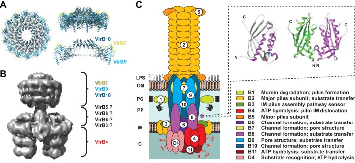
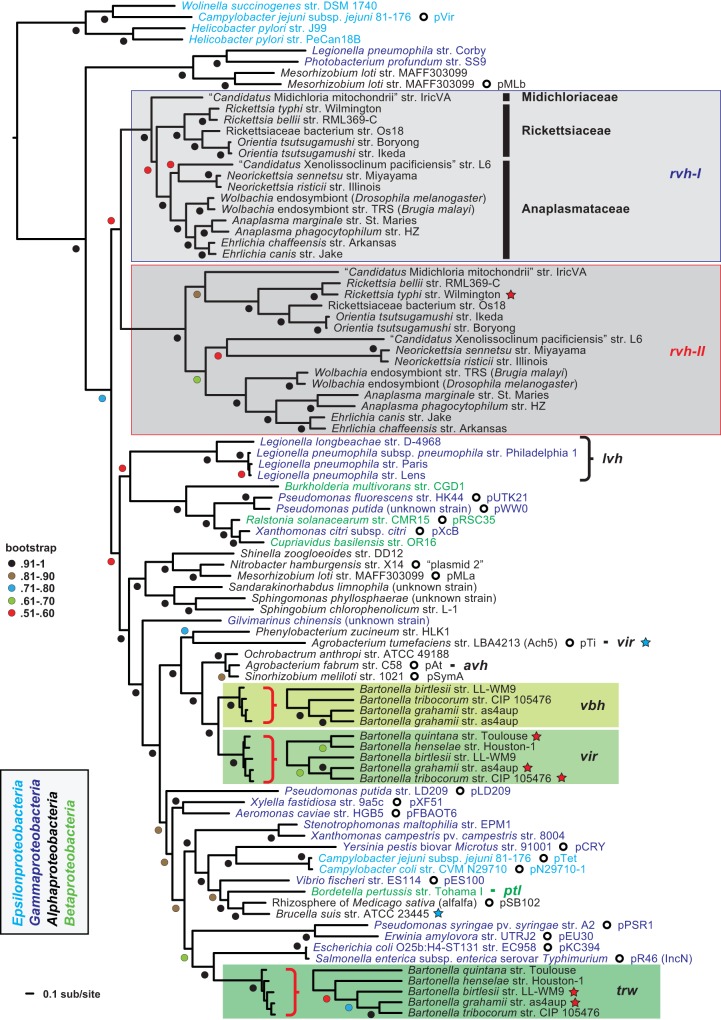
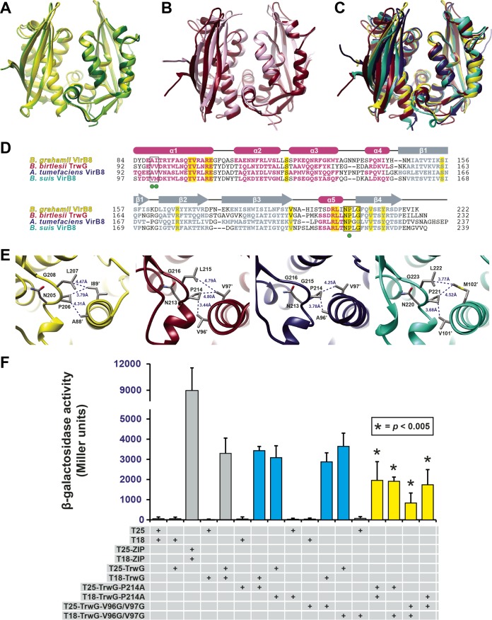
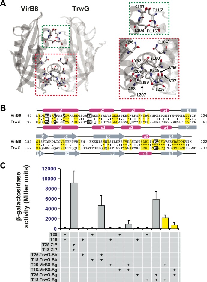
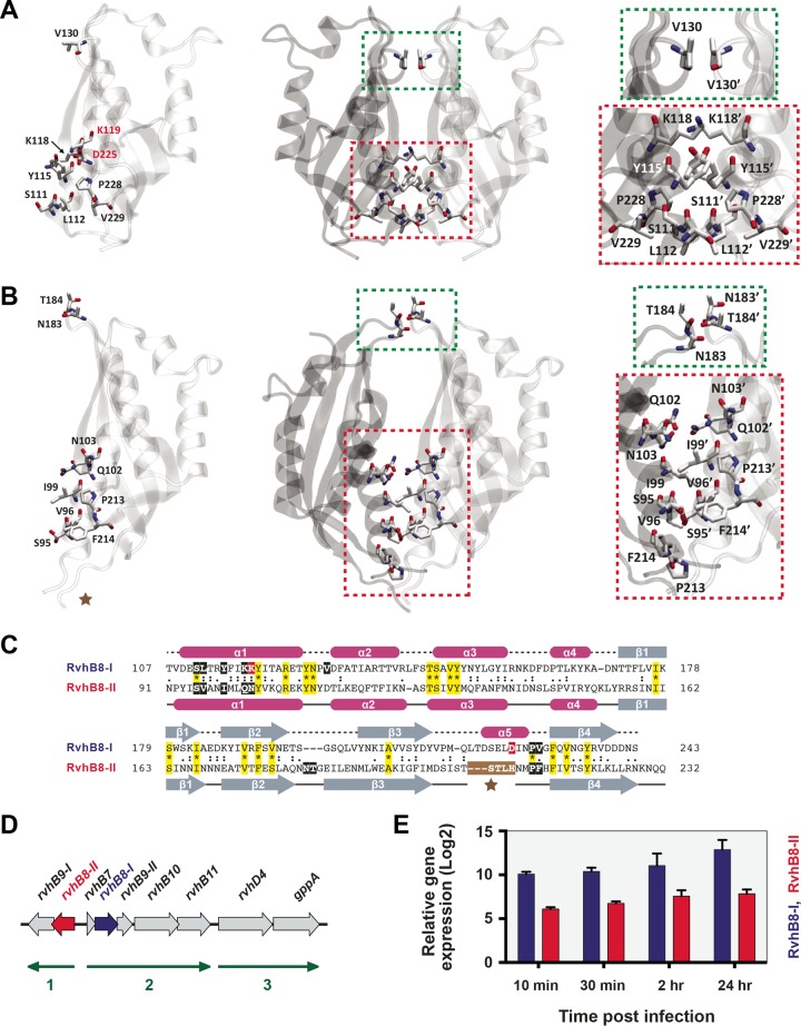
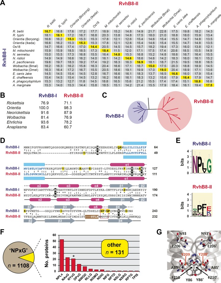
Prokaryotes use type IV secretion systems (T4SSs) to translocate substrates (e.g., nucleoprotein, DNA, and protein) and/or elaborate surface structures (i.e., pili or adhesins). Bacterial genomes may encode multiple T4SSs, e.g., there are three functionally divergent T4SSs in some Bartonella species (vir, vbh, and trw). In a unique case, most rickettsial species encode a T4SS (rvh) enriched with gene duplication. Within single genomes, the evolutionary and functional implications of cross-system interchangeability of analogous T4SS protein components remains poorly understood. To lend insight into cross-system interchangeability, we analyzed the VirB8 family of T4SS channel proteins. Crystal structures of three VirB8 and two TrwG Bartonella proteins revealed highly conserved C-terminal periplasmic domain folds and dimerization interfaces, despite tremendous sequence divergence. This implies remarkable structural constraints for VirB8 components in the assembly of a functional T4SS. VirB8/TrwG heterodimers, determined via bacterial two-hybrid assays and molecular modeling, indicate that differential expression of trw and vir systems is the likely barrier to VirB8-TrwG interchangeability. We also determined the crystal structure of Rickettsia typhi RvhB8-II and modeled its coexpressed divergent paralog RvhB8-I. Remarkably, while RvhB8-I dimerizes and is structurally similar to other VirB8 proteins, the RvhB8-II dimer interface deviates substantially from other VirB8 structures, potentially preventing RvhB8-I/RvhB8-II heterodimerization. For the rvh T4SS, the evolution of divergent VirB8 paralogs implies a functional diversification that is unknown in other T4SSs. Collectively, our data identify two different constraints (spatiotemporal for Bartonella trw and vir T4SSs and structural for rvh T4SSs) that mediate the functionality of multiple divergent T4SSs within a single bacterium.

Importance: Assembly of multiprotein complexes at the right time and at the right cellular location is a fundamentally important task for any organism. In this respect, bacteria that express multiple analogous type IV secretion systems (T4SSs), each composed of around 12 different components, face an overwhelming complexity. Our work here presents the first structural investigation on factors regulating the maintenance of multiple T4SSs within a single bacterium. The structural data imply that the T4SS-expressing bacteria rely on two strategies to prevent cross-system interchangeability: (i) tight temporal regulation of expression or (ii) rapid diversification of the T4SS components. T4SSs are ideal drug targets provided that no analogous counterparts are known from eukaryotes. Drugs targeting the barriers to cross-system interchangeability (i.e., regulators) could dysregulate the structural and functional independence of discrete systems, potentially creating interference that prevents their efficient coordination throughout bacterial infection.


Figures

FIG 1
FIG 1
Architecture of P-type type IV secretions systems (P-T4SSs). (A) Bird’s-eye (left) and side (right) views of the dodecameric P-T4SS core complex (CC) encoded by plasmid pKM101 of Escherichia coli (PDB ID 3JQO), adapted from the work of Chandran et al. (17). Colors for the CC subunits (VirB7, VirB9, and VirB10) are similar to the model in panel C. (B) Negative-station electron microscopy-generated structure of the P-T4SS encoded by the E. coli R388 conjugative plasmid (EMD-2567) (adapted from the work of Low et al. [19]). Colors for the CC subunits and cytosolic/IM barrels (VirB4) are similar to the model in panel C. The putative positions of other IM channel (IMC) proteins are depicted with questions marks. VirB1, VirB2, VirB11, and VirD4 are not shown, as they were not included in the original structure. (C) General model of the composition of P-T4SSs, with functions for all 12 components (VirB1 to VirB11 and VirD4) listed at bottom right. The purple star depicts the bitopic VirB8 IMC proteins, with a dashed box illustrating the monomeric (left) and dimeric (right) structures for VirB8 C-terminal domains of Agrobacterium tumefaciens (PDB ID 2CC3) (44).
FIG 2
FIG 2
Phylogeny estimation of P-type type IV secretion systems (P-T4SSs). Phylogeny was estimated from concatenated alignments of five components (VirB4 and VirB8 to VirB11), except for rvhB-II, which contains homologs to VirB4, VirB8, and VirB9 only (see the text for further details on alignment and data set construction). ML-based phylogeny was estimated with RAxML on the unmasked alignment (LG + gamma + Ι). Branch support was assessed with 1,000 bootstrap pseudoreplications. A tree with specific bootstrap values, as well as additional phylogenies estimated from alternative alignments/models/optimality criteria, is provided in Text S1 in the supplemental material. The tree was rooted with four P-T4SSs from species of Epsilonproteobacteria. rvh-I and rvh-II T4SSs are in light and dark gray, respectively. The Bartonella vir, vbh, and trw T4SSs are colored different shades of green. Red stars depict sequences used to generate VirB8 structures in this study; blue stars depict sequences previously used to determine VirB8 structures. Plasmid-encoded T4SSs are depicted with open circles, with plasmid names provided. NCBI GenBank accession numbers for all proteins are provided in Text S1 in the supplemental material.
FIG 3
FIG 3
Bartonella VirB8 and TrwG proteins form structurally conserved homodimers. (A) Superimposition of the ribbon representations of the VirB8 crystal structures from B. quintana strain Toulouse (dark green) (PDB ID 4LSO), B. grahamii strain as4aup (yellow) (PDB ID 4KZ1) and B. tribocorum strain CIP 105476 (light green) (PDB ID 4MEI). (B) Superimposition of the ribbon representations of the TrwG crystal structures from B. birtlesii strain LL-WM9 (burgundy) (PDB ID 4JF8) and B. grahamii strain as4aup (pink) (PDB ID 4NHF). (C) Superimposition of the ribbon representations for the crystal structures of B. grahamii VirB8, B. birtlesii TrwG, Agrobacterium tumefaciens strain C58 VirB8 (dark blue) (PDB ID 2CC3) (44), and Brucella suis biovar 1 (strain 1330) VirB8 (aquamarine) (PDB ID 2BHM) (45). (D) Sequence alignment of VirB8/TrwG proteins and secondary structure assignment. Sequences were extracted from a larger alignment (see Text S4 in the supplemental material). Sequences are the globular domains depicted in panel C. For each protein, residues involved in the major dimerization site are boxed. Invariant residues are highlighted in yellow. Magenta bars and gray arrows depict the α-helices and β-strands for the VirB8/TrwG structure, with colored residues in the proteins corresponding to these structural features. Green dots mark the residues mutated within the dimerization interface of B. birtlesii TrwG. (E) High-resolution depiction of the major dimerization sites for the proteins illustrated in panels C and D. From left to right: B. grahamii VirB8, B. birtlesii TrwG, A. tumefaciens VirB8, and B. suis VirB8. (F) Analysis of B. birtlesii TrwG-TrwG interactions using the bacterial two-hybrid system. Different plasmid combinations were transformed into adenylate cyclase deficient and cAMP-specific-phosphodiesterase-deficient E. coli strain APE304. After overnight growth in liquid Luria-Bertani medium, β-galactosidase activity was measured and calculated (in Miller units). T25-ZIP and T18-ZIP are positive-interaction control plasmids encoding dimer-forming yeast transcription factor GCN4 (89). Values are from three independent experiments, all analyzed in triplicate. Blue bars depict wild-type-mutant interactions, while yellow bars depict mutant-mutant interactions.
FIG 4
FIG 4
In vitro heterodimerization of Bartonella VirB8 and TrwG proteins. (A) Ribbon representation of a modeled heterodimer comprised of the VirB8 (PDB ID 4KZ1) and TrwG (PDB ID 4NHF) crystal structures from B. grahamii strain as4aup. Regions involved in dimerization are boxed and enlarged at right. Green indicates a minor dimerization site involving residues within the loop between α-helices α1 and α2. Red indicates a major dimerization site involving residues of α-helix α1 and the NPXG motif. TrwG residues are denoted with primes. (B) Sequence alignment of B. grahamii VirB8 and TrwG proteins, with secondary structure assignment. Sequences were extracted from a larger alignment (see Text S4 in the supplemental material). Sequences are the globular domains depicted in panel A. For each protein, residues involved in the dimerization interface are within black boxes. Invariant residues are highlighted in yellow. Magenta bars and gray arrows depict the α-helices and β-strands for the VirB8 and TrwG structures. (C) Analysis of B. birtlesii strain LL-WM9 and B. grahamii TrwG-TrwG interactions, the B. grahamii VirB8-VirB8 interaction, and the B. grahamii VirB8-TrwG interaction using the bacterial two-hybrid system. See the legend to Fig. 3F and the text for descriptions of the bacterial two-hybrid assay. Yellow bars depict the interactions tested for the B. grahamii VirB8-TrwG heterodimer.
FIG 5
FIG 5
Rickettsia typhi expresses two structurally divergent VirB8-like proteins. (A) Structural model for Rtyphi RvhB8-1 (RT0280; YP_067242). (Left) RvhB8-I monomer in ribbon representation with nine residues involved in the dimerization interface shown in stick representation. For clarity, residues colored red are not shown in the dimer. (Center) RvhB8-I dimer in ribbon representation with seven residues involved in the dimerization interface shown in stick representation. Green indicates a minor dimerization site involving residues within the loop between α-helices α1 and α2. Red indicates a major dimerization site involving residues of α-helix α1 and the NPXG motif. (Right) Higher magnification of the RvhB8-I subunit interface. (B) Ribbon representation for the monomer and dimer of the crystal structure (PDB ID 4O3V) of Rtyphi RvhB8-II (RT0278; YP_067240). Depiction of dimerization scheme follows the layout shown for RvhB8-I in panel A, except that the minor dimerization site (green) involved residues between β-strands β2 and β3. The brown star denotes the break in the structure. (C) Sequence alignment of RvhB8-I and RvhB8-II proteins and secondary structure assignment. Sequences are those of the globular domains depicted in panels A and B. For each protein, residues involved in the dimerization interface are within black (or red) boxes. Invariant residues are highlighted yellow. Magenta cylinders and gray arrows depict the α-helices and β-strands, respectively, for structures shown in panels A and B. For RvhB8-II, the brown star denotes the break in the structure, with missing residues colored white. (D) rvhB8-I and rvhB8-II are arrayed in tandem operons within the Rtyphi genome. The schema shows nucleotide coordinates 351539 to 360917 from the Rtyphi strain Wilmington genome (NC_006142) (46). Nine genes are encoded within three predicted operons: 1, rvhB9-I and rvhB8-II (red); 2, rvhB7, rvhB8-I (blue), rvhB9-II, rvhB10, and rvhB11; 3, rvhD4 and gppA (guanosine-5′-triphosphate,3′-diphosphate pyrophosphatase). The direction of transcription for each operon is shown with green arrows. Operons were predicted with fgenesb (90). Other rvh genes are encoded in separate clusters within the Rtyphi genome (41). (E) Expression of the Rtyphi rvhB8-I and rvhB8-II genes during early host cell infection. RNA was extracted from HeLa cells infected with R. typhi, and gene expression of RT0280 (encoding RvhB8-I) and RT0278 (encoding RvhB8-II) was measured by reverse transcription-quantitative PCR (RT-qPCR). Gene expression was normalized to R. typhi reference genes adr1 and sca5 (2ΔCT). Infections were repeated in triplicate with technical duplicate readings for RT-qPCR. Values are means and standard errors of the means.
FIG 6
FIG 6
Conserved RvhB8-I and RvhB8-II paralogs are highly divergent from one another. (A) Pairwise divergence between RvhB8-I and RvhB8-II proteins from select rickettsial species. Numbers are amino acid identity (percent) as calculated across a global RvhB8 alignment (see the text for details). Highlighted values on the diagonal depict divergences between paralogs encoded within the same genome. Full species names and NCBI GenBank accession numbers for all proteins are provided in Text S1 in the supplemental material. (B) Across species and strains within the same genus, RvhB8-I is more conserved than RvhB8-II. Numbers are amino acid identity (percent) as described for panel A. Complete percent identity matrices used to estimate protein divergence are provided in Text S5 in the supplemental material. (C) Phylogeny estimation of RvhB8 proteins reveals higher divergence within the RvhB8-II clade than the RvhB8-I clade. ML-based phylogeny was estimated with RAxML on the unmasked global RvhB8 alignment (WAG + gamma + Ι). A complete tree, as well as phylogenies estimated from the masked alignment with other substitution models, is provided in Text S5 supplemental material. (D) Comparison of Rtyphi RvhB8-I and RvhB8-II proteins. Sequences were aligned according to structure in SPDBV using “magic fit” followed by “improved fit” algorithms. The alignment shows predicted (top) and solved (bottom) structures for RvhB8-I and RvhB8-II, respectively. Predicted transmembrane-spanning regions (76) are colored blue. Five residues conserved across all RvhB8 proteins are in black, with residues conserved only in RvhB8-I (n = 15) or RvhB8-II (n = 1) highlighted in yellow (see Text S5 in the supplemental material). The NPXG motif is in a red box (see the description of panel E below). For RvhB8-II, the region of proteolysis (STLH) that occurred during crystallization is in a brown box. (E) Composition of the NPXG motif across 15 RvhB8-I (top) and 15 RvhB8-II (bottom) proteins. Sequence logos were generated using WebLogo v.3.3 (77). (F) Analysis of the conservation of the NPXG motif across 1,239 nonredundant proteobacterial VirB8 proteins (excluding Rickettsiales). Proteins lacking the conserved NPXG motif (10.6%) were placed in 14 categories based on their alternative sequences and ranked by their frequency (see Text S4 in the supplemental material for structural modeling of proteins within each category). (G) Example of a canonical interaction across the NPXG motif for Yersinia pestis biovar microtus strain 91001 (NP_995427), which contains the alternative sequence NYFG.

Similar articles

Cited by

References

    1. Alvarez-Martinez CE, Christie PJ. 2009. Biological diversity of prokaryotic type IV secretion systems. Microbiol Mol Biol Rev 73:775–808. doi:10.1128/MMBR.00023-09. - DOI - PMC - PubMed
    1. Christie PJ, Atmakuri K, Krishnamoorthy V, Jakubowski S, Cascales E. 2005. Biogenesis, architecture, and function of bacterial type IV secretion systems. Annu Rev Microbiol 59:451–485. doi:10.1146/annurev.micro.58.030603.123630. - DOI - PMC - PubMed
    1. Lawley TD, Klimke WA, Gubbins MJ, Frost LS. 2003. F factor conjugation is a true type IV secretion system. FEMS Microbiol Lett 224:1–15. doi:10.1016/S0378-1097(03)00430-0. - DOI - PubMed
    1. Hofreuter D, Odenbreit S, Haas R. 2001. Natural transformation competence in Helicobacter pylori is mediated by the basic components of a type IV secretion system. Mol Microbiol 41:379–391. doi:10.1046/j.1365-2958.2001.02502.x. - DOI - PubMed
    1. Hamilton HL, Domínguez NM, Schwartz KJ, Hackett KT, Dillard JP. 2005. Neisseria gonorrhoeae secretes chromosomal DNA via a novel type IV secretion system. Mol Microbiol 55:1704–1721. doi:10.1111/j.1365-2958.2005.04521.x. - DOI - PubMed

Publication types

Substances

LinkOut - more resources