. 2016 Apr;73(8):1715-39.
doi: 10.1007/s00018-015-2099-4. Epub 2015 Dec 8.

Endoglin regulates mural cell adhesion in the circulatory system

Affiliations

Endoglin regulates mural cell adhesion in the circulatory system

Elisa Rossi et al. Cell Mol Life Sci. 2016 Apr.

Abstract

The circulatory system is walled off by different cell types, including vascular mural cells and podocytes. The interaction and interplay between endothelial cells (ECs) and mural cells, such as vascular smooth muscle cells or pericytes, play a pivotal role in vascular biology. Endoglin is an RGD-containing counter-receptor for β1 integrins and is highly expressed by ECs during angiogenesis. We find that the adhesion between vascular ECs and mural cells is enhanced by integrin activators and inhibited upon suppression of membrane endoglin or β1-integrin, as well as by addition of soluble endoglin (SolEng), anti-integrin α5β1 antibody or an RGD peptide. Analysis of different endoglin mutants, allowed the mapping of the endoglin RGD motif as involved in the adhesion process. In Eng (+/-) mice, a model for hereditary hemorrhagic telangectasia type 1, endoglin haploinsufficiency induces a pericyte-dependent increase in vascular permeability. Also, transgenic mice overexpressing SolEng, an animal model for preeclampsia, show podocyturia, suggesting that SolEng is responsible for podocytes detachment from glomerular capillaries. These results suggest a critical role for endoglin in integrin-mediated adhesion of mural cells and provide a better understanding on the mechanisms of vessel maturation in normal physiology as well as in pathologies such as preeclampsia or hereditary hemorrhagic telangiectasia.

Keywords: Blood vessels; Cell adhesion; HHT; Kidney; TGF-β; Tubulogenesis.


Figures
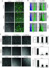
Fig. 1
Fig. 1
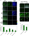
Endoglin silencing in endothelial and smooth muscle cells. a Primary cultures of HUVECs, HAECs and UASMCs (p4–8) were untreated (control), nucleofected with endoglin (Eng) specific siRNA (#s4677 and #s4679) and GFP (green stain), or nucleofected with scrambled siRNA (#AM4611 and #AM4613) and GFP (siRNA control). After 48 h, cells were observed by confocal microscopy. Cells cotransfected with GFP are visualized by their green fluorescence. b Flow cytometry analysis. Primary cultures of HUVECs, HAECs and UASMCs were untreated (control) or nucleofected with Eng siRNA or scrambled siRNA #AM4611 and #AM4613 (siRNA control). After 48 h, cells were analyzed by immunofluorescence flow cytometry with anti-endoglin mAb P4A4 (green histograms) or a negative control mAb (X63; blue histograms). The mean fluorescence intensity (MFI) of each sample is indicated. Endoglin expression is decreased upon transfection with specific siRNA in all cells. Cells nucleofected with scrambled siRNA showed the same endoglin expression levels as untreated cells (data not shown). c HUVECs and HAECs were incubated in matrigel to analyze tube formation. Confocal microscopy of untreated cells (control), nucleofected for 48 h with scrambled siRNA (AM4611, #1; AM4613, #2) or Eng siRNA (#s4677 and #s4679), or incubated with soluble endoglin (Sol.Eng) are shown. The histogram on the right indicates the percentage, respect to the control sample (100 %), of closing tubes under each experimental condition. Samples were in triplicates and the mean of the control condition was given the arbitrary value of 100. The average of five different experiments is shown. The statistical significance respect to control value (CTR) is indicated (***p < 0.001)
Fig. 2
Fig. 2
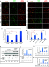
Role of endoglin in adhesion between VSMCs and ECs. a, b Cell–cell adhesion in angiogenesis assays. a UASMCs were transfected with GFP and cocultured with unlabeled HUVECs at a 1:4 ratio in matrigel to analyze mural cell adhesion to ECs. Confocal microscopy of untreated cells (control) and cells incubated with soluble endoglin (Sol.Eng) or nucleofected with Eng siRNA are shown in representative photographs. The intensity of the staining according to the color scale (0–250) indicates mural cell adhesion to endothelial cells in 3D co-culture b Quantification of UASMCs binding to ECs was carried out by measuring the intensity profile using fluorescence confocal microscopy (SP5, Leica). The mean area in percentage, representing mural cell adhesion measured in different fields, is indicated. Samples were in triplicates and the mean of the control condition was given the arbitrary value of 100. The average of five different experiments is shown. c, d Cell adhesion assay. c HUVEC monolayers were incubated with UASMCs previously labeled with CSFE in the absence or in the presence of soluble endoglin. After 1 h incubation, wells were washed and the cells were visualized by confocal microscopy. d Binding of UASMCs to HUVECs in c was quantified by measuring the intensity profile using fluorescence confocal microscopy (SP5, Leica). The average of four independent experiments is shown. The statistical significance respect to control value (CTR) is indicated. *p < 0.05; **p < 0.005; ***p < 0.001
Fig. 3
Fig. 3
Role of integrins in adhesion between VSMCs and ECs. UASMCs were labeled with CSFE (green) and HUVECs were labeled with CMTPX (red). Then, UASMCs were cocultured with HUVECs at a 1:4 ratio in matrigel to analyze mural cell adhesion to ECs. a, c Representative photographs of confocal microscopy analysis. a Cells treated with a control mAb IgG2b (CTR) and cells incubated with the inhibitory anti-β1 integrins mAb LIA1/2, the anti-β1 integrins mAb TS2/16 or the general integrins activator MnCl2. c Untreated cells (control) and cells incubated with the pericyte recruiter PDGF-BB or the integrins activator CXCL12 either in the absence or in the presence of the chemokine receptor (CXCR4) inhibitor AMD3100 (AMD). b, d Quantification of UASMCs binding to HUVECs from a and c, respectively. The intensity profile was measured using fluorescence confocal microscopy (SP5, Leica). The mean area in percentage, representing mural cell adhesion measured in different fields, is indicated. Histograms in b and d represent the mean of four and five independent experiments, respectively. The statistical significance respect to control value (CTR) is indicated. *p < 0.05; **p < 0.005; ns not significant. eh Effect of soluble endoglin on Akt and FAK phosphorylation. UASMCs were transfected or not with β1-integrin siRNA or scramble siRNA (scRNA). Cultures of UASMCs or cocultures of UASMCs and HAECs were incubated in the absence or presence of 1 μg/mL SolEng. At the times indicated, adherent cells were lysed and proteins were subjected to SDS-PAGE, followed by immunodetection with anti-p-FAK (Tyr925), anti-pAkt (Ser473) or anti-actin antibodies (e). Histograms represent the p-FAK/actin ratio in UASMCs (f), p-FAK/actin ratio in UASMCs/HAECs (g) and the p-Akt/actin ratio in UASMCs/HAECs (h). This is a representative experiment of five different ones
Fig. 4
Fig. 4
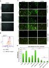
Silencing of β1-integrin in UASMCs. Primary cultures of UASMCs were transfected with beta1 integrin specific siRNA (siRNA-β1) or scrambled siRNA (scRNA). Transfected UASMCs were morphologically (a), phenotypically (b) and functionally (c, d) analyzed. a Untreated UASMCs (control) and cells transfected scRNA display the same morphology with slight changes respect to cells transfected with siRNA-β1, likely due to the β1 integrin role in cell adhesion. b Immunofluorescence flow cytometry with anti-CD29 (anti-β1 integrin) antibodies show a downregulation of β1 integrin (76 %) in UASMCs transfected with specific siRNA vs. cells transfected with scrambled siRNA. c Cell–cell adhesion assays. Confluent monolayers of HAECs were incubated with UASMCs, previously labeled with CFSE, in the absence (control) or presence of 1 μg/mL SolEng or 100 ng/mL CXCL12, as indicated. After 1 h incubation, wells were washed and the cells were visualized by confocal microscopy. d Binding of HAECs to UASMCs in c was quantified by measuring the fluorescence intensity using Image J and Histolab™ (Microvision) software. A representative experiment out of four made in triplicate with similar results is shown (**p < 0.005; ***p < 0.001)
Fig. 5
Fig. 5
Involvement of the endoglin RGD motif in adhesion between VSMCs and ECs. a HUVECs, labeled with CSFE (green), were incubated with the UASMC monolayers for 1 h at 37 °C with/without thalidomide, CXCL12 (CXC), SolEng (S.Eng), RGD peptide or DGR peptide, as indicated. Bound CSFE-labeled cells were lysed and quantified by Varioskan plate reader. The average of four different experiments in duplicate is shown. Statistical significances vs. control cells (**p < 0.005) or vs. CXCL12-treated cells (**p < 0.005; red asterisks) are indicated. be Adhesion of different deletion constructs of endoglin to VSMCs. c Atomic model for endoglin and location of RGD motif. The extracellular domain of endoglin contains an orphan domain (OD) and a juxtamembrane zona pellucida domain (ZPD) that includes an RGD motif (in magenta). The dashed line separates the OD from the ZPD. d Generation of different truncated forms of endoglin. Numbers indicate the amino acid of endoglin (starting at the N terminus) that limit the corresponding fragment. The position of extracellular (EC), transmembrane (TM), and cytoplasmic (CT) domains, is indicated. All of the constructs contain the leader sequence of the IgGκ and the HA epitope at the N terminus (from the pDisplay vector), and construct 437/586-Endo encode the transmembrane domain of the pDisplay vector. The OD encompasses amino acid residues 26–359, whereas the ZP domain is contained within the fragment 360–586. The ZPD-C (residues 437–586) is a sub-domain of ZPD. The presence of the RGD motif (residues 399–401) and its mutant version (RGA) is indicated. b, e Adhesion assays of Jurkat cells to VSMCs. Jurkat cells were nucleofected with endoglin, ZPD-Endo, ZPD-Endo-RGA or 437/586-Endo constructs together with GFP. As negative controls, cells were nucleofected with GFP in the absence (CTR) or in the presence of the empty vector (EV). After nucleofection, Jurkat cells were added to the UASMC monolayers for 1 h at 37 °C. Bound GFP expressing cells were lysed and quantified by Varioskan plate reader (e). A representative experiment out of three made in triplicate with similar results is shown. Statistical significances vs. control cells (**p < 0.005) or cells expressing full length endoglin (*p < 0.05; red asterisks) are indicated
Fig. 6
Fig. 6
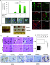
Soluble endoglin binds to VSMCs and inhibits angiogenesis in vivo. a Binding of soluble endoglin to VSMCs. Exponentially growing UASMCs were incubated with a chimeric protein containing the extracellular domain of endoglin fused to the Fc fragment of IgG (Sol.Eng-Fc), and the integrins’ activators CXCL12 and MnCl2, as indicated. After incubation with FITC-labeled anti-human IgG, cells were fixed, counterstained with DAPI and analyzed by confocal microscopy (lower panel). The fluorescence intensity was measured using confocal microscopy (SP5, Leica) and represented as mean values per cell. A representative experiment out of three made in triplicate with similar results is shown. The statistical significance is indicated (**p < 0.005). b In vitro 7 days co-culture of bmMPC and ECFC. bmMPC differentiate into VSMC/pericytes as evidenced by the positivity for the antibodies specific for αSMA, PDGFRβ, NG2, Calponin I and Sm22α (green stain). A negative control antibody (IgG) was also used. Endothelial cells (ECFC) are stained in red using an antibody against von Willebrand factor. cf Effect of soluble endoglin on in vivo angiogenesis. c Nude mice (n = 3) were injected with the coculture of bmMPC + ECFC in matrigel either in the absence (control) or in the presence of soluble endoglin (Sol.Eng; 5 μg/mL/plug). d After 1 week, plugs were extracted from the animals. e Plug sections were stained with hematoxylin and eosin. Arrowheads indicate the presence of vascular structures containing erythrocytes. f Quantification of the vessels. The average of three different experiments made in triplicate is shown (N nude mice = 9; n = 9 plugs for each condition). The plugs treated with SolEng display a lower number of vessels compared to controls (*p < 0.04). g Immunohistochemistry of plug sections. Arrowheads indicate the staining of vascular endothelium with anti-CD31 antibodies and mural cells with anti-αSMA. Staining of αSMA is almost absent in plugs treated with soluble endoglin. A positive staining of arterioles from the neighboring tissue is included as an internal control
Fig. 7
Fig. 7
Effect of silencing endoglin and β1 integrin in angiogenesis assays in vivo. a Suppression of β1 integrin and endoglin using specific siRNA was carried out in cultured bmMPC and ECFC, respectively. Endothelial cells (ECFC) or smooth muscle cells (bmMPC9) were stained with anti-CD105 (endoglin) in green or anti-CD29 (β1 integrin) in red, respectively. b Quantification of the vessels. The average of two different experiments performed in triplicate is shown. The individual or combined suppression of endoglin or β1 integrin in bmMPC or ECFC, respectively, leads to a markedly reduced number of vessels compared to controls (**p < 0.01; ***p < 0.005). c Four groups of nude mice (n = 3 each) were injected with the coculture of bmMPC + ECFC in matrigel either in the absence (scrambled siRNA; control) or in the presence of siRNA-mediated suppression, as indicated. After 1 week, plugs were extracted from the animals and stained with hematoxylin and eosin. Arrowheads indicate the presence of vascular structures containing erythrocytes
Fig. 8
Fig. 8
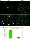
Increased vascular permeability in Eng +/− mice. Eng +/− and Eng +/+ mice were perfused through the jugular vein with FITC-dextran. After 2 h, animals were killed and eyes were removed and dissected. Nineteen neuroretinas from ten different animals (six Eng +/+ and four Eng +/− mice) were isolated and mounted with antifade solution. The green fluorescent labeling of the retinas was visualized using a fluorescence microscope (Axiovert 200M, Zeiss). a Representative photographs from three Eng +/+ and three Eng +/− mice, are shown. An increased number of permeability foci and diffuse green fluorescence background is observed in Eng +/− mice (arrows) compared with Eng +/+. b Quantification of permeability spots (**p = 0.01)
Fig. 9
Fig. 9
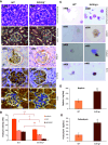
Analysis of podocytes in the kidney from Sol.Eng + mice. a Comparison between glomeruli of Sol.Eng + mice (n = 17) and controls (n = 17). The overall structure of glomeruli in WT and transgenic animals is similar, as evidenced by the hematoxylin and eosin and trichromic staining, although a certain degree of leukocyte infiltration was observed in Sol.Eng + mice. Immunostaining with anti-αSMA, anti-podocin and anti-WT1 shows a positive signal in podocytes from wild type animals and a reduced staining in the Sol.Eng + glomeruli. Representative tissue sections are shown. b The immunostaining of αSMA, podocin and WT1 shown in a was quantified using the Image J software. Relative units (RU) of podocyte staining from four different experiments are represented in the vertical axis. c Thinprep on urine from Sol.Eng + mice (n = 5) and controls (n = 5). Hematoxylin and eosin staining shows the presence of cells in urine. Staining with anti-α-SMA, anti-podocin and anti-WT1 indicates the presence of podocytes in the urine of Sol.Eng + mice. d, g Protein levels of nephrin and podocalyxin were measured in the urine of WT (n = 5) and Sol.Eng + (n = 5) mice using an ELISA kit
Fig. 10
Fig. 10
Schematic diagram showing the role of endoglin in integrin-mediated cell adhesion between ECs and mural cells/podocytes. ac Blood vessels. a A normal blood vessel with an endothelial monolayer facing the lumen surrounded by vascular mural cells (VMCs) and ECM proteins. ECs and vascular mural cells share a common basal membrane (BM). During vascular development and stabilization, binding of the homeostatic chemokine CXCL12 to its receptor CXCR4 leads to activation of β1-integrins in VMCs. Then, endothelial endoglin binds to β1-integrins on VMCs. b In HHT1, endoglin haploinsufficiency leads to a decreased binding of endothelial endoglin to β1-integrins in VMCs. c In preeclampsia, soluble endoglin competes with membrane bound endoglin for the binding to β1-integrins in VMCs. d, e Kidney glomerulus. d A normal glomerulus showing pericytes bound to the glomerular basal membrane (GBM) through their surface integrins. e In preeclampsia, soluble endoglin competes with GBM for the binding to surface integrins in podocytes. The presence of endothelial endoglin in the lumen of the vessel and the existence of other adhesion molecules have been omitted for simplification

References

    1. Armulik A, Genove G, Betsholtz C. Pericytes: developmental, physiological, and pathological perspectives, problems, and promises. Dev Cell. 2011;21:193–215. doi: 10.1016/j.devcel.2011.07.001. - DOI - PubMed
    1. Davis GE, Stratman AN, Sacharidou A, Koh W. Molecular basis for endothelial lumen formation and tubulogenesis during vasculogenesis and angiogenic sprouting. Int Rev Cell Mol Biol. 2011;288:101–165. doi: 10.1016/B978-0-12-386041-5.00003-0. - DOI - PMC - PubMed
    1. Stratman AN, Davis GE. Endothelial cell–pericyte interactions stimulate basement membrane matrix assembly: influence on vascular tube remodeling, maturation, and stabilization. Microsc Microanal. 2012;18:68–80. doi: 10.1017/S1431927611012402. - DOI - PMC - PubMed
    1. Aguilera KY, Brekken RA. Recruitment and retention: factors that affect pericyte migration. Cell Mol Life Sci. 2014;71:299–309. doi: 10.1007/s00018-013-1432-z. - DOI - PMC - PubMed
    1. Marcelo KL, Goldie LC, Hirschi KK. Regulation of endothelial cell differentiation and specification. Circ Res. 2013;112:1272–1287. doi: 10.1161/CIRCRESAHA.113.300506. - DOI - PMC - PubMed

Publication types

MeSH terms