. 2016 Jul;44(7):e509-19.
doi: 10.1097/CCM.0000000000001475.

Short-Term Dietary Restriction Rescues Mice From Lethal Abdominal Sepsis and Endotoxemia and Reduces the Inflammatory/Coagulant Potential of Adipose Tissue

Affiliations

Short-Term Dietary Restriction Rescues Mice From Lethal Abdominal Sepsis and Endotoxemia and Reduces the Inflammatory/Coagulant Potential of Adipose Tissue

Marlene E Starr et al. Crit Care Med. 2016 Jul.

Abstract

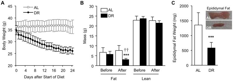
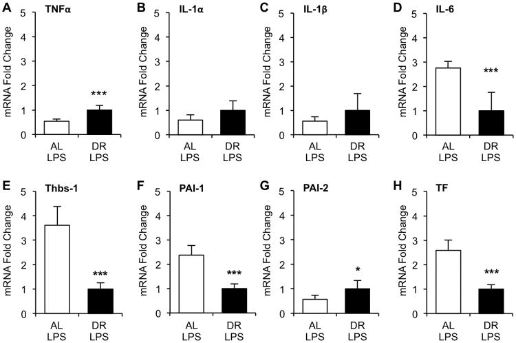
Objectives: Visceral adipose tissue is a major site for expression of proinflammatory and procoagulant genes during acute systemic inflammation. In this study, we tested whether the loss of fat mass by dietary restriction would remove the major source of these factors resulting in improved tolerance to sepsis and endotoxemia.

Design: Prospective, laboratory controlled experiments.

Setting: Aging and critical care research laboratory in a university hospital.

Subjects: Middle-aged (12-month old) male C57BL/6 mice.

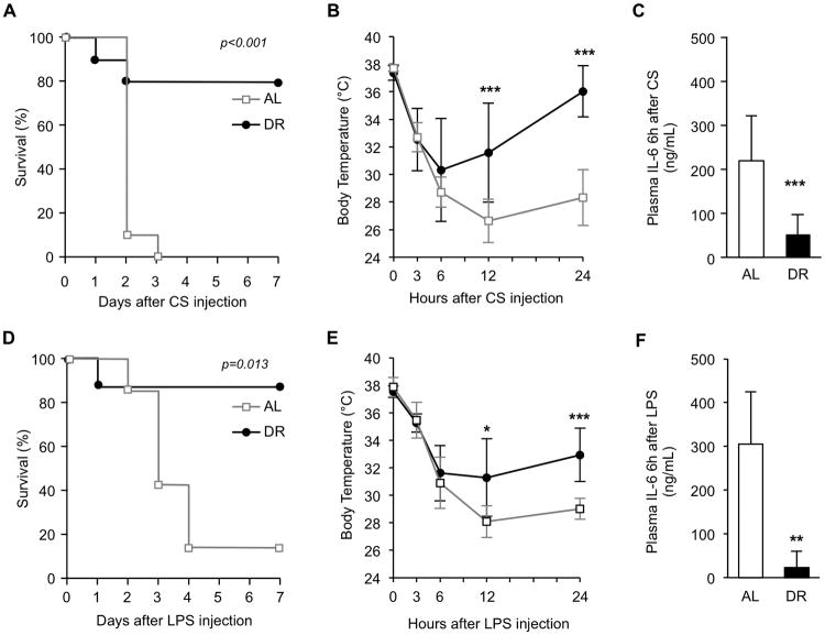
Interventions: Mice were subjected to 40% dietary restriction for 3 weeks followed by induction of abdominal sepsis or endotoxemia by intraperitoneal injection with cecal slurry or lipopolysaccharide, respectively.

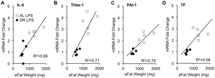
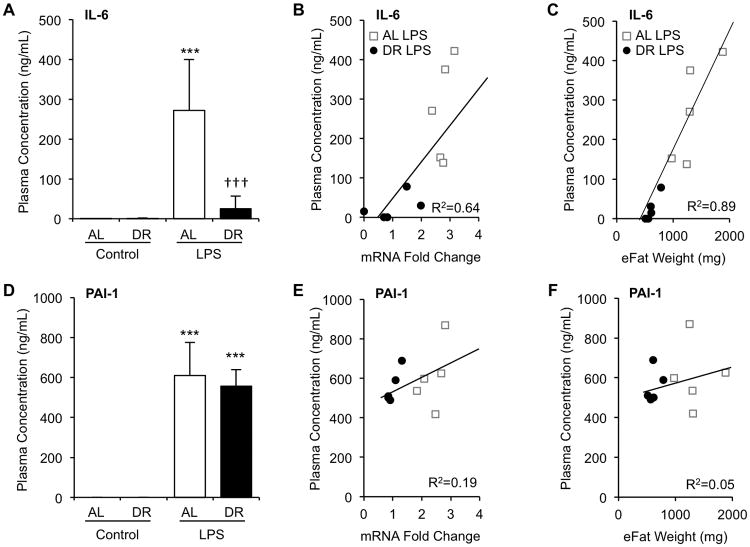
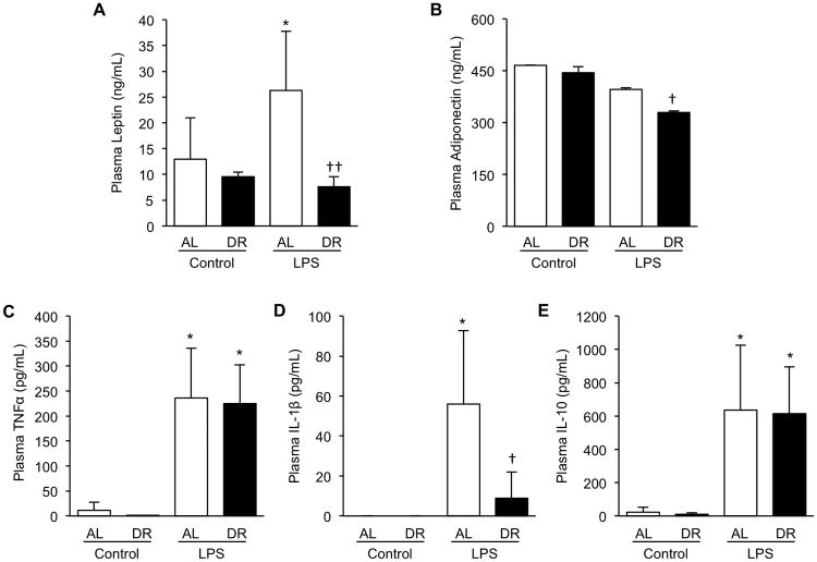
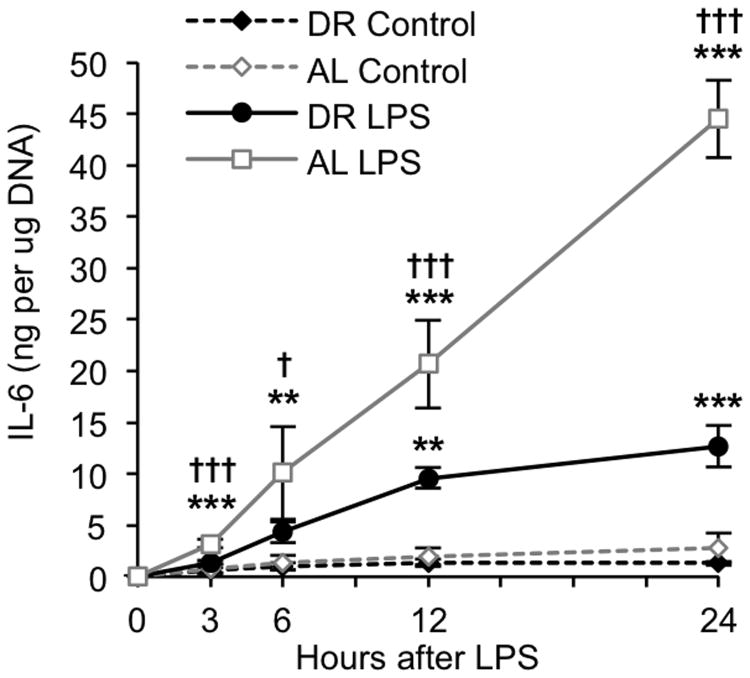
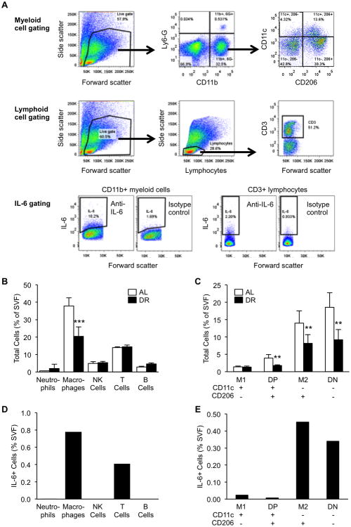
Measurements and main results: Compared with freely fed mice, dietary restricted mice exhibited dramatically improved survival (80% vs 0% after sepsis; p < 0.001 and 86% vs 12% after endotoxemia; p = 0.013) and significantly reduced visceral fat-derived messenger RNA expression of interleukin-6, thrombospondin-1, plasminogen activator inhibitor-1, and tissue factor, which positively correlated with fat mass. Plasma levels of interleukin-6 were significantly reduced by dietary restriction and correlated with adipose interleukin-6 messenger RNA levels and fat mass (p < 0.001; R = 0.64 and 0.89). In vitro culture of visceral fat explants from naive dietary restricted mice showed significantly reduced interleukin-6 secretion compared with that from freely fed mice in response to lipopolysaccharide. Analysis of major adipose immune cell populations by flow cytometry demonstrated that macrophages were the only cell population reduced by dietary restriction and that CD11c/CD206 (M2-type) and CD11c/CD206 (double negative) macrophages, in addition to T cells, are the major immune cell populations that produce interleukin-6 in middle-aged mice during systemic inflammation.

Conclusions: Short-term dietary restriction drastically improved the survival outcome of middle-aged mice during both polymicrobial sepsis and sterile endotoxemia. Improved survival was accompanied by a significantly attenuated inflammatory response in adipose tissue, which is likely due to alterations of both fat mass quantity and qualitative changes, including a reduction in macrophage populations.


Conflict of interest statement

Copyright form disclosures: Drs. Saito, Starr, and Steele received support for article research from the National Institutes of Health (NIH). Dr. Cohen disclosed that he does not have any potential conflicts of interest.

Figures

Figure 1
Figure 1
Analysis of body weight and body composition in ad libitum (AL) fed mice and mice under short-term 40% dietary restriction (DR). (A) Daily body weight measurements of DR (n=8) and AL (n=8) mice during the DR period (prior to sepsis/endotoxemia induction). (B) Fat and lean mass weight before and after DR assessed by Echo MRI. (C) Total epididymal fat weight at sacrifice and representative macroscopic images. * indicates statistical significance compared to pre-DR measurements, † indicates statistical significance compared to AL group. Two and three symbols indicate p< 0.01 and 0.001, respectively.
Figure 2
Figure 2
Effects of short-term dietary restriction (DR) on survival during polymicrobial sepsis and endotoxemia. (A) After three weeks of DR (n=10) or ad libitum feeding (AL) (n=10), mice were injected with cecal slurry (CS, 200 μL, i.p) and survival monitored for one week. (B) Body temperature during the first 24h of sepsis. (C) Plasma IL-6 concentration, 6h after CS injection. (D) After three weeks of DR (n=8) or AL-feeding (n=7), mice were injected with LPS (2.5mg/kg, i.p) and survival monitored for one week. (E) Body temperature during the first 24h of endotoxemia. (F) Plasma IL-6 concentration, 6h after LPS injection. Values are the mean ± SD. * indicates statistical significance compared to AL group. One, two, and three symbols indicate p<0.05, 0.01, and 0.001, respectively.
Figure 3
Figure 3
Expression of inflammatory and coagulant genes in adipose tissue of dietary restricted (DR) and ad libitum (AL)-fed mice during endotoxemia. After three weeks of DR, DR and AL-fed mice (n=5 each) were injected with LPS (2.5mg/kg, i.p.) and sacrificed 6h later. Epididymal adipose tissues were harvested and extracted RNA processed and subjected to real-time qRT-PCR analysis for expression of pro-inflammatory factors (A) TNFα, (B) IL-1α, (C) IL-1β, (D)IL-6, and pro-coagulant factors (E) thrombospondin-1 (Thbs-1), (F) plasminogen activator inhibitor (PAI)-1, (G) PAI-2, (H) tissue factor (TF). Values are the mean ± SD, * indicates statistical significance compared to AL group. One and three symbols indicate p< 0.05 and 0.001, respectively.
Figure 4
Figure 4
Correlation between fat mass and adipose-derived gene expression among genes which are downregulated by dietary restriction (DR). Gene expression of (A) IL-6, (B) Thbs-1, (C) PAI-1, and (D) TF from epididymal adipose tissues (eFat) were plotted against the total weight of both fat pads.
Figure 5
Figure 5
Plasma IL-6, not PAI-1, correlates with adipose tissue gene expression and adiposity. After three weeks of DR, mice were injected with LPS (2.5mg/kg, i.p.) and sacrificed 6h later. Plasma was obtained by centrifugation of heparinized blood drawn from the inferior vena cava and subjected to analysis by ELISA. (A) Plasma IL-6 concentration. (B) Correlation of plasma IL-6 concentration with mRNA level of IL-6. (C) Correlation of plasma IL-6 concentration with weight of epididymal fat pads (eFat). (D) Plasma PAI-1 concentration. (E) Correlation of plasma PAI-1 concentration with mRNA level of PAI-1. (F) Correlation of plasma PAI-1 concentration with weight of eFat pads. Values are the mean ± SD, n=3 for non-injected controls, n=5 for LPS injected animals, * indicates statistical significance compared to non-injected controls of the same treatment group, † indicates statistical significance compared to AL LPS group. Three symbols indicate p< 0.001.
Figure 6
Figure 6
Plasma protein analysis of dietary restricted (DR) and ad libitum (AL)-fed mice during endotoxemia. After three weeks of DR, mice were injected with LPS (2.5mg/kg, i.p.) and sacrificed 6h later. Plasma was obtained by centrifugation of heparinized blood drawn from the inferior vena cava and subjected to analysis by ELISA. (A) Plasma adiponectin concentration, (B) Plasma leptin concentration, (C) Plasma TNFα concentration, (D) Plasma IL-1β concentration, (E) Plasma IL-10 concentration. Values are the mean ± SD, n=3 for non-injected controls, n=5 for LPS injected animals, * indicates statistical significance compared to non-injected controls of the same treatment group, † indicates statistical significance compared to AL LPS group. One and two symbols indicate p< 0.05 and 0.01, respectively.
Figure 7
Figure 7
Effects of dietary restriction (DR) on IL-6 release from in vitro cultured adipose tissues. Explant cultures of epididymal adipose tissue from naïve DR and AL-fed mice were treated with LPS (10μg/mL) in vitro and the medium sampled at multiple time-points for analysis of IL-6 by ELISA. Cytokine levels were adjusted for adipose tissue DNA content. Data are expressed as the mean ± SD, n=3 per group and time-point. * indicates a statistically significant change as compared to the 0h time-point of the same group. † indicates a statistically significant difference between DR and AL at the same time-point. One, two, and three symbols indicate p<0.05, 0.01, and 0.001, respectively.
Figure 8
Figure 8
Flow cytometric analysis of dietary restriction (DR)-mediated changes in adipose immune cell populations and identification of IL-6 producing cell types. (A) Gating strategy for delineation of myeloid cells and lymphoid cells. (B-C) Cell populations were quantified as a percentage of total SVF cells obtained from AL and DR mice. Data are expressed as the mean ± SD, n=5 per group and time-point. * indicates a statistically significant change as compared to the AL group. Two and three symbols indicate p<0.01 and 0.001, respectively. (D-E) IL-6 positive cells were quantified as a percentage of total SVF cells after adjusting for isotype control expression. Data are derived from SVF cells pooled from multiple AL-fed mice. IL-6 positive cells were not detected in NK or B cells.

References

    1. Starr ME, Hu Y, Stromberg AJ, et al. Gene expression profile of mouse white adipose tissue during inflammatory stress: age-dependent upregulation of major procoagulant factors. Aging cell. 2013;12(2):194–206. - PMC - PubMed
    1. Masoro EJ. Overview of caloric restriction and ageing. Mechanisms of ageing and development. 2005;126(9):913–922. - PubMed
    1. McCay CM, Crowell MF, Maynard LA. The effect of retarded growth upon the length of life span and upon the ultimate body size. 1935. J Nutrition. 1935;10:63–79. - PubMed
    1. Masoro EJ. Hormesis and the antiaging action of dietary restriction. Experimental gerontology. 1998;33(1-2):61–66. - PubMed
    1. Robertson LT, Mitchell JR. Benefits of short-term dietary restriction in mammals. Experimental gerontology. 2013;48(10):1043–1048. - PMC - PubMed

Publication types