. 2015 Dec 8;10(12):e0143484.
doi: 10.1371/journal.pone.0143484. eCollection 2015.

Interleukin-13 Inhibits Lipopolysaccharide-Induced BPIFA1 Expression in Nasal Epithelial Cells

Affiliations

Interleukin-13 Inhibits Lipopolysaccharide-Induced BPIFA1 Expression in Nasal Epithelial Cells

Yung-An Tsou et al. PLoS One. .

Abstract

Short palate, lung, and nasal epithelium clone 1 (SPLUNC1) protein is expressed in human nasopharyngeal and respiratory epithelium and has demonstrated antimicrobial activity. SPLUNC1 is now referred to as bactericidal/permeability-increasing fold containing family A, member 1 (BPIFA1). Reduced BPIFA1 expression is associated with bacterial colonization in patients with chronic rhinosinusitis with nasal polyps (CRSwNP). Interleukin 13 (IL-13), predominately secreted by T helper 2 (TH2) cells, has been found to contribute to airway allergies and suppress BPIFA1 expression in nasal epithelial cells. However, the molecular mechanism of IL-13 perturbation of bacterial infection and BPIFA1 expression in host airways remains unclear. In this study, we found that lipopolysaccharide (LPS)-induced BPIFA1 expression in nasal epithelial cells was mediated through the JNK/c-Jun signaling pathway and AP-1 activation. We further demonstrated that IL-13 downregulated the LPS-induced activation of phosphorylated JNK and c-Jun, followed by attenuation of BPIFA1 expression. Moreover, the immunohistochemical analysis showed that IL-13 prominently suppressed BPIFA1 expression in eosinophilic CRSwNP patients with bacterial infection. Taken together, these results suggest that IL-13 plays a critical role in attenuation of bacteria-induced BPIFA1 expression that may result in eosinophilic CRSwNP.


Conflict of interest statement

Competing Interests: The authors have declared that no competing interests exist.

Figures

Fig 1
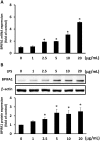
Fig 1. LPS induces BPIFA1 expression in nasal epithelial cells.
(A) RPMI-2650 cells were treated with various concentrations (0–20 μg/ml) of LPS for 2 h, and mRNA levels of BPIFA1 were analyzed by quantitative real-time PCR. (B) Cell lysates were then prepared to measure BPIFA1 protein expression levels by western blot after incubation with LPS for 24 h. Protein expression levels of BPIFA1 were quantified by densitometric analysis and normalized to those of β-actin. ANOVA with Tukey’s test was used to compare the overall difference between the groups. *, P < 0.05 compared to LPS-untreated control group.
Fig 2
Fig 2. Phosphorylated p65 does not respond to LPS-mediated BPIFA1 expression.
(A) RPMI-2650 cells were treated with LPS (10 μg/ml) for the indicated times, and the expression levels of phosphorylated ERK, JNK, p38, and p65 were determined by western blot. β-actin was used as the loading control. (B) Protein expression levels were quantified with densitometric analysis and presented as mean ± standard deviation, derived from three independent experiments.
Fig 3
Fig 3. JNK/c-Jun pathway is involved in LPS-mediated up-regulation of BPIFA1 expression in nasal epithelial cells.
(A) Cells were pretreated for 1 h with 20 μM PD98059 (ERK inhibitor), 10 μM SP600125 (JNK inhibitor), or 20 μM SB203580 (p38 inhibitor) and incubated with 10 μg/ml LPS for 2 h. (B) Cells were transfected with a JNK-dominant negative (DN-JNK) mutant for 24 h or pretreated with SP600125 for 30 min prior to incubation with LPS for 1 h. The protein expression levels were determined using western blot. β-actin was used as the loading control. The western blots were carried out independently in triplicate and results were representative of one of three independent experiments. The expression level of each protein was quantified by signal intensity and was indicated at the bottom of each lane. The quantitative analysis of western blot for three independent experiments was shown in S2 Fig. ANOVA with Tukey’s test was used to compare the overall difference between the groups. P < 0.05 was considered statistically significant.
Fig 4
Fig 4. AP-1 is involved in LPS-induced BPIFA1 expression.
Cells were pretreated with 10 μM curcumin or tanshinone (inhibitors of c-Jun) for 30 min, followed by incubation with LPS (10 μg/ml) for 2 h. Protein expression levels were determined using western blot and normalized to those of β-actin. The expression level of each protein is quantified by signal intensity and is indicated at the bottom of each lane. The quantitative analysis of western blot for three independent experiments was shown in S3 Fig. ANOVA with Tukey’s test was used to compare the overall difference between the groups. P < 0.05 was considered statistically significant. (B) Cells were transfected with AP-1-Luc reporter and incubated with LPS (10 μg/ml) for another 2 h. Cell lysates were subjected to luciferase activity assays to determine AP-1 luciferase activity. The results represented mean and standard deviation values from three independent experiments. *, P < 0.05 compared between two groups.
Fig 5
Fig 5. Inhibition of c-Jun suppresses LPS-induced BPIFA1 expression.
Cells were pretreated with 10 μM of SP600125, curcumin, or tanshinone (inhibitors of c-Jun) for 30 min, followed by incubation with LPS (10 μg/ml) for 2 h. Cells were washed and treated with antibodies against (A) phosphorylated c-Jun or (B) BPIFA1, followed by incubation with FITC–conjugated anti-mouse IgG (green). Cells were probed with DAPI to visualize the nucleus (blue) and analyzed by confocal fluorescence microscopy. Bars, 10 μm.
Fig 6
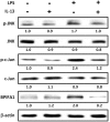
Fig 6. IL-13-mediates attenuation of LPS-induced BPIFA1 expression.
RPMI-2650 cells, either treated or untreated with IL-13 (10 ng/ml) for 48 h, were incubated with or without LPS (10 μg/ml) for further 2 h. Cell lysates were prepared to analyze the protein expression levels by using western blot. β-actin was used as the loading control. The western blots were carried out independently in triplicate and results were representative of one of three independent experiments. The expression level of each protein was quantified by signal intensity and was indicated at the bottom of each lane. The quantitative analysis of western blot for three independent experiments was shown in S4 Fig. ANOVA with Tukey’s test was used to compare the overall difference between the groups. P < 0.05 was considered statistically significant.
Fig 7
Fig 7. Bacterial infection is associated with elevated IL-13 secretion and reduced BPIFA1 expression in sinonasal tissues.
Representative immunohistochemical staining of BPIFA1 (upper panel) and IL-13 (lower panel) expressions in eosinophilic CRSwNP patients (A) without or (B, C) with bacterial infection. Arrows indicated the expression of IL-13 in the bacterial infection tissue (C). The images were photographed at a magnification of 200×.
Fig 8
Fig 8. Model depicting IL-13 inhibition of LPS-induced BPIFA1 expression in nasal epithelial cells.
(A) The bacterial cell wall component LPS upregulates BPIFA1 expression through the JNK/c-Jun signaling pathway, followed by AP-1 activation. (B) IL-13, a TH2-skewed cytokine, suppresses LPS-induced BPIFA1 expression in nasal epithelial cells from patients with eosinophilic CRSwNP.

References

    1. Di YP, Harper R, Zhao Y, Pahlavan N, Finkbeiner W, Wu R. Molecular cloning and characterization of spurt, a human novel gene that is retinoic acid-inducible and encodes a secretory protein specific in upper respiratory tracts. J Biol Chem 2003;278: 1165–1173. - PubMed
    1. Bingle CD, Gorr SU. Host defense in oral and airway epithelia: chromosome 20 contributes a new protein family. Int J Biochem Cell Biol 2004;36: 2144–2152. - PubMed
    1. Bingle CD, Seal RL, Craven CJ. Systematic nomenclature for the PLUNC/PSP/BSP30/SMGB proteins as a subfamily of the BPI fold-containing superfamily. Biochem Soc Trans 2011;39: 977–983. 10.1042/BST0390977 - DOI - PMC - PubMed
    1. Di YP. Functional roles of SPLUNC1 in the innate immune response against Gram-negative bacteria. Biochem Soc Trans 2007;39: 1051–1055. - PMC - PubMed
    1. Lukinskiene L, Liu Y, Reynolds SD, Steele C, Stripp BR, Leikauf GD, et al. Antimicrobial activity of PLUNC protects against Pseudomonas aeruginosa infection. J Immunol 2011;187: 382–390. 10.4049/jimmunol.1001769 - DOI - PMC - PubMed

Publication types

MeSH terms