. 2015 Dec 15;6(40):42575-89.
doi: 10.18632/oncotarget.6471.

Hormone stimulation of androgen receptor mediates dynamic changes in DNA methylation patterns at regulatory elements

Affiliations

Hormone stimulation of androgen receptor mediates dynamic changes in DNA methylation patterns at regulatory elements

Vineet K Dhiman et al. Oncotarget. .

Abstract

DNA methylation is an epigenetic modification that contributes to stable gene silencing by interfering with the ability of transcriptional regulators to bind to DNA. Recent findings have revealed that hormone stimulation of certain nuclear receptors induces rapid, dynamic changes in DNA methylation patterns alongside transcriptional responses at a subset of target loci, over time. However, the ability of androgen receptor (AR) to dynamically regulate gene transcription is relatively under-studied and its role in the regulation of DNA methylation patterns remains to be elucidated. Here we demonstrate in normal prostate cells that hormone stimulated AR activity results in dynamic changes in the transcription rate and DNA methylation patterns at the AR target genes, TIPARP and SGK1. Time-resolved chromatin immunoprecipitation experiments on the SGK1 locus reveals dynamic recruitment of AR and RNA Polymerase II, as well as the recruitment of proteins involved in the DNA demethylation process, TET1 and TDG. Furthermore, the presence of DNA methylation at dynamic regions inhibits protein binding and transcriptional activity of SGK1. These findings establish AR activity as a contributing factor to the dynamic regulation of DNA methylation patterns at target genes in prostate biology and infer further complexity involved in nuclear receptor mediation of transcriptional regulation.

Keywords: Chromosome Section; DNA methylation; androgen receptor; androgen regulated target genes; gene transcription; nuclear receptor.


Conflict of interest statement

CONFLICTS OF INTEREST

The authors declare that they have no conflicts of interest with the contents of this article.

Figures
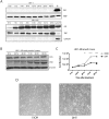
Figure 1
Figure 1. Characterization of short-term androgen treatment of HPr1-AR cell lines
A. HPr1 (upper panel) and HPr1-AR cells (lower panel) were treated with 10 nM DHT. Nuclear lysates were harvested at indicated time points for western blot analysis for AR and TBP. Positive control (+) represents HPr1-AR nuclear lysate stimulated with DHT for 48 h. Cyto AR represents the cytoplasmic fraction for AR, stimulated with DHT for 48 h. B. Cells were treated with 10 nM DHT are harvested every 24 h for total protein. Lysates were used in western blot analysis to determine differentiation potential of HPr1-AR cells. C. Cellular proliferation was determined using trypan blue staining. HPr1-AR cells were treated with 10 nM DHT and counted every 24 h. D. Microscopy representation of HPr1-AR cells after 48 h of EtOH vehicle or 10 nM DHT treatment. (Student T-test, **p < 0.01, ****p < 0.0001).
Figure 2
Figure 2. Bovine pituitary extract removal reduces nuclear AR levels and results in more reproducible transcript levels
A. Bovine pituitary extract (BPE) was removed from the growth media and nuclear lysates were collected every 24 h. B. HPr1-AR cells were maintained in either full or BPE-removed media conditions and then treated with EtOH vehicle or 10 nM DHT for 0 h, 2 h or 4 h. Total mRNA expression was analyzed via qRT-PCR. Error bars of indicative of standard error. (Student T-test, *p < 0.05).
Figure 3
Figure 3. Androgen-induced transcriptional output correlates with dynamic changes in the DNA methylation patterns of gene regulatory elements
A. Schematic representation of the SGK1 (upper panel) and TIPARP (lower panel) loci. Outlined in gray boxes are the ARE regions interrogated for methylation analysis. B.-C. HPr-1AR cells were treated with 10nM DHT and harvested for RNA and DNA, from the same cell pellet, at the indicated time points. Transcription data is reflective of normalized quantity of nascent gene expression. Expression data was normalized to β-microglobulin and representative of the mean of three biological replicates. CpG sites in the TIPARP ARE II and SGK1 ARE I regions were interrogated for methylation. Each row represents a single CpG site, where each CpG site is labeled in respect to its distance in base pairs from the TSS (as noted by the arrow). The black box represents the confirmed androgen response element (ARE) from previously published ChIP-chip studies. [24] All methylation data points are representative of the mean of three biological replicates or best two out of three replicates. Error bars of indicative of standard error.
Figure 4
Figure 4. Dynamic recruitment of AR, TDG and TET1 is observed at the SGK1 ARE I
HPr-1AR cells were treated with EtOH vehicle or 10 nM DHT for indicated time points. Regulatory element occupancy was determined using antibodies against A. AR, B. TDG and C. TET1 and normalized and presented as fold change over input. ChIP signal was measured using qRT-PCR with primers specific to the interrogated region. All data points are representative of biological triplicates. Error bars are indicative of standard error. (Student T-test, *p < 0.05).
Figure 5
Figure 5. Methylation of dynamic CpG sites at the SGK1 ARE I inhibit the binding of protein to DNA
A. Schematic representation of the SGK1 locus. An in-depth sequence analysis of the ARE I region illustrates 12 different CpG sites (highlighted in red). The ARE I region contains two AR binding sequences (as boxed), where one binding site is representative of a full consensus sequence and the second site is representative of a half-site. Designed DNA probes for EMSA analysis are highlighted in yellow. Probe A consists of CpG sites −1024 and −1029. Probe B consists of CpG site −1079. B. DNA probes were either unmethylated or methylated, radiolabeled, and were incubated with DHT-stimulated HPr1-AR nuclear extract, an unlabeled competitor probe and/or and unlabeled methylated competitor probe.
Figure 6
Figure 6. Methylation of the SGK1 ARE I region inhibits transcriptional activity
The SGK1 ARE I region was cloned into the pCpGfree-promoter vector. Unmethylated, partially methylated (HpaII) and completely methylated (SssI) versions of the vector were transfected into HPr1-AR cells. Cells were treated with EtOH vehicle or 10 nM DHT for 48 h and assayed for Lucia (RLU) and secreted alkaline phosphatase (SEAP) expression. All data points represent three biological replicates. (Student T-test, *p < 0.05).

References

    1. Evans RM. The steroid and thyroid hormone receptor superfamily. Science. 1988;240:889–895. - PMC - PubMed
    1. Helsen C, Claessens F. Looking at nuclear receptors from a new angle. Mol Cell Endocrinol. 2014;382:97–106. - PubMed
    1. Glass C, Rosenfeld M. The coregulator exchange in transcriptional functions of nuclear receptors. Genes Dev. 2000;14:22. - PubMed
    1. Carlberg C, Seuter S. Dynamics of nuclear receptor target gene regulation. Chromosoma. 2010;119:479–484. - PMC - PubMed
    1. Biddie SC, Hager GL. Glucocorticoid receptor dynamics and gene regulation. Stress. 2009;12:193–205. - PMC - PubMed

Publication types

MeSH terms