. 2015 Dec 8;11(12):e1005324.
doi: 10.1371/journal.ppat.1005324. eCollection 2015 Dec.

Characterization of a Novel Human-Specific STING Agonist that Elicits Antiviral Activity Against Emerging Alphaviruses

Affiliations

Characterization of a Novel Human-Specific STING Agonist that Elicits Antiviral Activity Against Emerging Alphaviruses

Tina M Sali et al. PLoS Pathog. .

Abstract

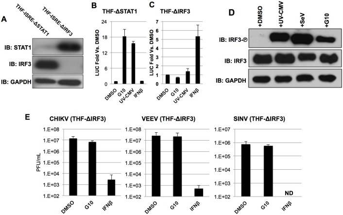
Pharmacologic stimulation of innate immune processes represents an attractive strategy to achieve multiple therapeutic outcomes including inhibition of virus replication, boosting antitumor immunity, and enhancing vaccine immunogenicity. In light of this we sought to identify small molecules capable of activating the type I interferon (IFN) response by way of the transcription factor IFN regulatory factor 3 (IRF3). A high throughput in vitro screen yielded 4-(2-chloro-6-fluorobenzyl)-N-(furan-2-ylmethyl)-3-oxo-3,4-dihydro-2H-benzo[b][1,4]thiazine-6-carboxamide (referred to herein as G10), which was found to trigger IRF3/IFN-associated transcription in human fibroblasts. Further examination of the cellular response to this molecule revealed expression of multiple IRF3-dependent antiviral effector genes as well as type I and III IFN subtypes. This led to the establishment of a cellular state that prevented replication of emerging Alphavirus species including Chikungunya virus, Venezuelan Equine Encephalitis virus, and Sindbis virus. To define cellular proteins essential to elicitation of the antiviral activity by the compound we employed a reverse genetics approach that utilized genome editing via CRISPR/Cas9 technology. This allowed the identification of IRF3, the IRF3-activating adaptor molecule STING, and the IFN-associated transcription factor STAT1 as required for observed gene induction and antiviral effects. Biochemical analysis indicates that G10 does not bind to STING directly, however. Thus the compound may represent the first synthetic small molecule characterized as an indirect activator of human STING-dependent phenotypes. In vivo stimulation of STING-dependent activity by an unrelated small molecule in a mouse model of Chikungunya virus infection blocked viremia demonstrating that pharmacologic activation of this signaling pathway may represent a feasible strategy for combating emerging Alphaviruses.


Conflict of interest statement

The authors have declared that no competing interests exist.

Figures

Fig 1
Fig 1. High Throughput Identification of G10. A. Luciferase (LUC) expression of top tier candidate molecules as described in text from a high throughput screen of 51,632 compounds.
(A) Presented values represent the average ±SD of duplicate assays. Black bar and arrow indicate LUC signal generated by G10. (B) Chemical structure of 4-(2-chloro-6-fluorobenzyl)-N-(furan-2-ylmethyl)-3-oxo-3,4-dihydro-2H-benzo[b][1,4]thiazine-6-carboxamide (G10).
Fig 2
Fig 2. G10 Induces IFN/IRF3- but not NF-κB-Dependent Transcription in Human Fibroblasts.
(A) Dose-dependent expression of IRF3/IFN-dependent luciferase (LUC) in telomerized human fibroblasts (THF). Values displayed are average fold changes ±SD of quadruplicate measurements of luminescence following 7h exposure to indicated concentration of G10 relative to cells exposed only to 1% DMSO (all samples normalized to 1% DMSO). (B) mRNA transcription of genes dependent on IRF3/IFN following 7h exposure to 100μM G10 or UV-CMV. Indicated values represent average ±SEM mRNA fold change relative to cells exposed to 1% DMSO from duplicate experiments. (C) Induction of NF-κB-dependent LUC signal in THF reporter cells following 7h exposure to 1μg/mL LPS, 160 HA units/mL SeV, 1ng/mL TNFα or indicated concentration of G10. Values displayed are as described in (A). (D) mRNA transcription of of NF-κB-dependent genes following 7h exposure to 100μM G10 or 160 HA units/mL SeV. Indicated values are mRNA fold change relative to cells exposed to 1% DMSO and are representative of duplicate experiments.
Fig 3
Fig 3. G10 Elicits Antiviral Activity.
Average titers ±SD of VACV, WNV, CHIKV, and VEEV grown on THF cells in the presence of indicated G10 concentration (DMSO concentration normalized to 1%). Infections were performed in triplicate and virus harvested at 48h post infection (CHIKV, WNV, CHIKV) or 24h post infection (VACV, VEEV).
Fig 4
Fig 4. IRF3 is Required for G10-Dependent Transcription and Anti-Alphaviral Activity.
(A) Immunoblot showing IRF3, STAT1, and GAPDH in THF-ISRE stably transduced with Cas9 and CRISPR gRNA directed against either STAT1 (THF-ISRE-ΔSTAT1) or IRF3 (THF-ISRE-ΔIRF3) as indicated. (B) Induction of IRF3/IFN-dependent LUC in THF lacking STAT1 following 7h exposure to 100μM G10, UV-inactivated CMV, or 1000U/mL IFNβ. Values displayed are average fold changes ±SD of quadruplicate measurements relative to cells exposed only to 1% DMSO. (C) Induction of IRF3/IFN-dependent LUC in THF lacking IRF3 following 7h exposure to 100μM G10, UV-inactivated CMV, or 1000U/mL IFNβ. Values displayed are as in B. (D) Immunoblot of lysates from THF-ISRE following 6h exposure to DMSO, UV-CMV, SeV or 100uM G10 as indicated showing phosphorylation status of IRF3 S386, total IRF3, and GAPDH. (E) Average media titers +SD of CHIKV, VEEV, and SINV at 24 (VEEV) or 48hpi (CHIKV, SINV) obtained from THF-ISRE-ΔIRF3 cells treated with 1% DMSO, 100μM G10, or 1000U/mL IFNβ as indicated. Infections were performed in triplicate.
Fig 5
Fig 5. G10 Elicits IRF3 phosphorylation and Anti-Alphaviral Activity in Cells Lacking IPS1.
(A) Immunoblot of lysates from THF-ISRE-ΔIPS1 following 6h exposure to DMSO, UV-CMV, SeV or 100uM G10 as indicated showing phosphorylation status of IRF3 S386, total IRF3, IPS1, STING, and GAPDH. (B) Average media titers +SD of CHIKV, VEEV, and SINV at 24h (VEEV) or 48h (CHIKV, SINV) post infection obtained from THF-ISRE-ΔIPS1 cells treated with 1% DMSO, 100μM G10, or 1000U/mL IFNβ as indicated. Infections were performed in triplicate.
Fig 6
Fig 6. G10-Mediated IRF3 Phosphorylation, ISG mRNA Induction, and Anti-Alphaviral Activity are not Detectable in Cells Lacking STING.
(A) Immunoblot of lysates from THF-ΔSTING following 7h exposure to 1% DMSO, 0.1μg/mL poly(I:C), SeV, UV-CMV, 1μg/mL 2’3’-cGAMP, or 100uM G10 as indicated showing phosphorylation status of IRF3 S386, total IRF3, IPS1, STING, and GAPDH. (B) Expression of IRF3/IFN-dependent LUC in THF-ISRE-ΔIPS1 and THF-ISRE-ΔSTING following 7h exposure to 1% DMSO, indicated concentrations of G10, SeV, UV-CMV, or 1μg/mL LPS. Values are presented as average fold change in quadruplicate measurements ±SD relative to cells treated with 1% DMSO. (C) Average fold changes ±SD from duplicate experiments of ISG54, ISG15, and Viperin mRNA relative to cells treated with 1% DMSO in THF-ISRE-ΔIPS1 (black bars) or THF-ISRE-ΔSTING (gray bars) following exposure to UV-CMV, SeV, or 100μM G10. (D) Media titers of CHIKV and VEEV at 24h (VEEV) or 48h (CHIKV) obtained from THF-ISRE-ΔSTING cells treated with 1% DMSO, 100μM G10, or 1000U/mL IFNβ as indicated. Infections were performed in triplicate.
Fig 7
Fig 7. G10-Mediated Expression of Type I and III IFN and IFN-Dependent Genes.
Induction of IFNβ and IFNλ1 (A) or Mx2 and OAS (B) transcripts in THF following 18h exposure to SeV, UV-CMV, or 100μM G10 or IFNβ as indicated. Values presented are average fold changes relative to cells treated with 1% DMSO ±SD based on duplicate treatments. (C) Secretion of type I IFN from THF-ISRE, THF-ISRE-ΔIPS1, or THF-ISRE-ΔSTING following 18h exposure to 1% DMSO, SeV, UV-CMV, or 100μM G10. Average LUC values ±SD were obtained from THF-ISRE-ΔIRF3 exposed (in triplicate) to media harvested from indicated cells and exposed to indicated stimulus (in triplicate).
Fig 8
Fig 8. Abrogation of G10-Mediated Antiviral Activity and ISG Expression in Cells Lacking STAT1.
(A) Average media titers ±SD of CHIKV, VEEV, and SINV at 24h (VEEV) or 48h (CHIKV, SINV) obtained from THF-ISRE-ΔSTAT1 cells treated with 1% DMSO, 100μM G10, or 1000U/mL IFNβ as indicated. Infections were performed in triplicate. (B) Synthesis of Mx2 and ISG56 proteins in THF-ISRE and THF-ISRE-ΔSTAT1 following 24h exposure to SeV, UV-CMV, 1000U/mL IFNβ or 100μM G10 as indicated.
Fig 9
Fig 9. Comparative Kinetics and Dose-Dependence of Innate Immune Activation by G10 and 2’3’-cGAMP.
(A) Immunoblot of lysates from THF-ISRE cells following exposure to G10 (100μM) or transfected 2’3’-cGAMP (42.3μM) for indicated time showing phosphorylation status of IRF3 S386, total IRF3, and GAPDH. (B) mRNA synthesis of indicated genes in THF following 8h exposure to indicated concentration of G10 (blue) or 2’3’-cGAMP (red). Indicated values represent average mRNA fold change ±SD from duplicate experiments relative to cells exposed to 1% DMSO.
Fig 10
Fig 10. G10-mediated Induction of mRNA in PBMCs.
mRNA synthesis of indicated genes in human peripheral blood mononuclear cells (PBMC) following 8h exposure to indicated concentration of G10 (A) or ppp-dsRNA (12.5μg/mL) or 2’3’-cGAMP (28μM) (B). Indicated values represent average mRNA fold change ±SD from duplicate experiments relative to cells exposed to 1% DMSO.
Fig 11
Fig 11. Innate Immune Activation by and Antiviral Activity of DMXAA.
A. Immunoblot of lysates from murine RAW264.7 cells following exposure to 100μM DMXAA for indicated time showing phosphorylation status of IRF3 S386, total IRF3, and GAPDH. (B) mRNA synthesis of indicated genes in RAW264.7 cells following 8h exposure to indicated concentration of DMXAA. Values represent average mRNA fold change ±SD from duplicate experiments relative to cells exposed to 1% DMSO. (B) Levels of serum-associated CHIKV at 72h post infection. Five mice per group were treated with DMSO alone or DMXAA at 3h pre- or 6h or 24h post inoculation as indicated.

References

    1. Broz P, Monack DM. Newly described pattern recognition receptors team up against intracellular pathogens. Nature Publishing Group. 2013;13: 551–565. 10.1038/nri3479 - DOI - PubMed
    1. Es-Saad S, Tremblay N, Baril M, Lamarre D. Regulators of innate immunity as novel targets for panviral therapeutics. Current Opinion in Virology. 2012;2: 622–628. 10.1016/j.coviro.2012.08.009 - DOI - PMC - PubMed
    1. Sharma S, tenOever BR, Grandvaux N, Zhou G-P, Lin R, Hiscott J. Triggering the interferon antiviral response through an IKK-related pathway. Science. American Association for the Advancement of Science; 2003;300: 1148–1151. 10.1126/science.1081315 - DOI - PubMed
    1. Yamamoto M, Sato S, Hemmi H, Hoshino K, Kaisho T, Sanjo H, et al. Role of adaptor TRIF in the MyD88-independent toll-like receptor signaling pathway. Science. American Association for the Advancement of Science; 2003;301: 640–643. 10.1126/science.1087262 - DOI - PubMed
    1. Oshiumi H, Matsumoto M, Funami K, Akazawa T, Seya T. TICAM-1, an adaptor molecule that participates in Toll-like receptor 3-mediated interferon-beta induction. Nat Immunol. 2003;4: 161–167. 10.1038/ni886 - DOI - PubMed

Publication types

MeSH terms